 Back
BackAutonomic Nervous System: Structure, Function, and Regulation
Study Guide - Smart Notes
 Tailored notes based on your materials, expanded with key definitions, examples, and context.
Autonomic Nervous System (ANS)
Overview and Function
The Autonomic Nervous System (ANS) is a division of the peripheral nervous system responsible for regulating involuntary functions of smooth muscle, cardiac muscle, and glands. It operates largely below the level of conscious awareness, ensuring optimal support for body activities such as heart rate, digestion, respiratory rate, pupillary response, urination, and sexual arousal.
Motor Neurons: ANS consists of motor neurons that innervate smooth and cardiac muscle, and glands.
Subconscious Control: Functions are regulated involuntarily, often referred to as the involuntary nervous system or general visceral motor system.
Homeostasis: The ANS maintains internal balance by adjusting physiological processes.
Structural Organization of the Nervous System
Somatic vs. Autonomic Nervous Systems
Both the somatic and autonomic nervous systems contain motor fibers, but they differ in their effectors, pathways, and responses to neurotransmitters.
Somatic Nervous System: Controls voluntary movements via skeletal muscles.
Autonomic Nervous System: Controls involuntary functions via cardiac muscle, smooth muscle, and glands.
Efferent Pathways:
Somatic: Single, thick myelinated axon from CNS to skeletal muscle.
Autonomic: Two-neuron chain (preganglionic and postganglionic neurons) from CNS to effector organs.
Neurotransmitter Effects:
Somatic: Always stimulatory (acetylcholine).
Autonomic: Can be stimulatory or inhibitory (acetylcholine or norepinephrine), depending on receptor type.
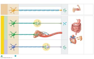
Divisions of the ANS
Sympathetic and Parasympathetic Divisions
The ANS is divided into two main branches: the sympathetic and parasympathetic divisions. Most visceral organs are innervated by both divisions, which often have opposing effects, maintaining dynamic antagonism and homeostasis.
Sympathetic Division: Mobilizes the body for activity ("fight-or-flight" response).
Parasympathetic Division: Promotes maintenance activities and conserves energy ("rest-and-digest" response).
Key Anatomical Differences
There are three main anatomical differences between the sympathetic and parasympathetic divisions:
Sites of Origin: Parasympathetic fibers originate in the brain and sacral spinal cord; sympathetic fibers originate in the thoracic and lumbar spinal cord.
Relative Lengths of Fibers: Parasympathetic has long preganglionic and short postganglionic fibers; sympathetic has short preganglionic and long postganglionic fibers.
Location of Ganglia: Parasympathetic ganglia are near or within effector organs; sympathetic ganglia are close to the spinal cord.
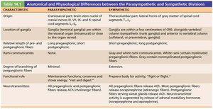
Parasympathetic Division
Craniosacral Origin and Pathways
The parasympathetic division is also known as the craniosacral division, as its fibers originate from the brainstem and sacral regions. Long preganglionic fibers extend from the CNS almost to target organs, synapsing in terminal ganglia close to or within the organs.
Cranial Nerves: Oculomotor (III), Facial (VII), Glossopharyngeal (IX), and Vagus (X) nerves carry parasympathetic fibers to various targets.
Functions: Directs digestion, diuresis, and defecation; maintains low heart rate and blood pressure.
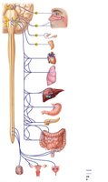
Sympathetic Division
Thoracolumbar Origin and Pathways
The sympathetic division is more complex and innervates more organs. Its fibers originate from the thoracic and lumbar regions of the spinal cord. Preganglionic neurons are located in spinal cord segments T1–L2 and synapse in ganglia near the spinal cord.
Functions: Mobilizes the body during activity, increases heart rate, dilates bronchioles, and releases glucose from the liver.
Unique Roles: Only sympathetic fibers innervate sweat glands, arrector pili muscles, kidneys, and most blood vessels.
Neurotransmitters and Receptors
Cholinergic and Adrenergic Fibers
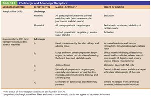
The major neurotransmitters of the ANS are acetylcholine (ACh) and norepinephrine (NE). The effects depend on the type of receptor present on the target organ.
Cholinergic Fibers: Release ACh; all ANS preganglionic axons and all parasympathetic postganglionic axons.
Adrenergic Fibers: Release NE; most sympathetic postganglionic axons.
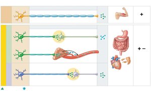
Cholinergic Receptors
Nicotinic Receptors: Found on skeletal muscle cells, all postganglionic neurons, and adrenal medulla; always excitatory.
Muscarinic Receptors: Found on all effector cells stimulated by postganglionic cholinergic fibers; can be excitatory or inhibitory depending on the target organ.
Adrenergic Receptors
Alpha (α) Receptors: Subclasses α1 and α2; effects depend on location.
Beta (β) Receptors: Subclasses β1, β2, and β3; effects depend on location.
Effects of Drugs on the ANS
Drug Classes and Clinical Applications
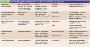
Various drugs influence the ANS by acting on its receptors. These include agents that stimulate or inhibit cholinergic and adrenergic receptors, affecting physiological responses.
Drug Class | Receptor Bound | Effects | Example | Clinical Application |
|---|---|---|---|---|
Nicotinic agents | Nicotinic ACh receptors | Stimulates sympathetic effects | Nicotine | Smoking cessation products |
Parasympathomimetic agents | Muscarinic ACh receptors | Enhances parasympathetic activity | Pilocarpine | Glaucoma treatment |
Acetylcholinesterase inhibitors | Inhibits breakdown of ACh | Enhances parasympathetic activity | Neostigmine | Treatment of myasthenia gravis |
Sympathomimetic agents | Adrenergic receptors | Enhances sympathetic activity | Albuterol | Treats asthma |
Sympatholytic agents | Adrenergic receptors | Decreases sympathetic activity | Propranolol | Treats hypertension |
Effects of Parasympathetic and Sympathetic Divisions on Organs
Organ-Specific Responses
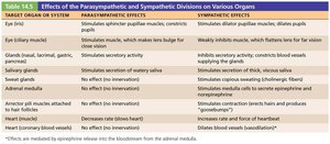
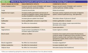
The parasympathetic and sympathetic divisions exert different effects on various organs, often antagonistic but sometimes cooperative.
Target Organ/System | Parasympathetic Effects | Sympathetic Effects |
|---|---|---|
Eye (iris) | Constricts pupil | Dilates pupil |
Heart | Decreases heart rate | Increases heart rate |
Lungs | Constricts bronchioles | Dilates bronchioles |
Digestive tract | Increases motility and secretion | Decreases motility and secretion |
Blood vessels | Little or no effect | Constriction and blood pressure regulation |
Control of ANS Function
CNS Centers and Regulation
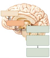
The ANS is regulated by several CNS centers, including the brain stem, spinal cord, hypothalamus, and cerebral cortex. The hypothalamus is the main integrative center, with input from the limbic system and cerebral cortex influencing autonomic responses.
Brain Stem: Directly influences heart rate, blood vessel diameter, and gastrointestinal activities.
Hypothalamus: Controls heart activity, blood pressure, body temperature, water balance, and endocrine activity.
Cerebral Cortex: Can modify ANS activity subconsciously, often through emotional responses.
Homeostatic Imbalances of the ANS
Common Disorders
Disorders of the ANS often involve deficient control of smooth muscle activity, leading to various clinical conditions:
Hypertension: Overactive sympathetic response increases blood pressure, treated with adrenergic receptor-blocking drugs.
Raynaud’s Disease: Exaggerated vasoconstriction in fingers and toes, treated with vasodilators.
Autonomic Dysreflexia: Uncontrolled activation of autonomic neurons in individuals with spinal cord injuries, leading to dangerous increases in blood pressure.
