 Back
Autonomic Regulation of the Eye, Auditory System, Muscle Structure, and Cardiac Physiology
Study Guide - Smart Notes
 Tailored notes based on your materials, expanded with key definitions, examples, and context.
Autonomic Regulation of the Eye
Pupillary Control: Parasympathetic and Sympathetic Pathways
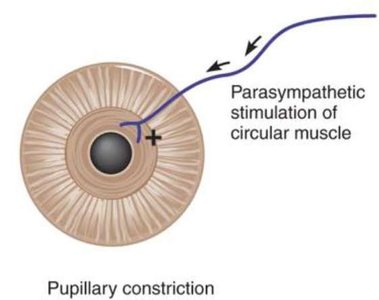
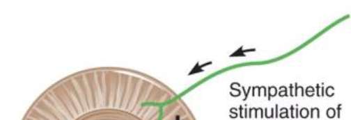
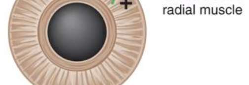
The diameter of the pupil is regulated by the autonomic nervous system, which controls two sets of smooth muscles in the iris: the circular (sphincter) and radial (dilator) muscles. These muscles adjust the amount of light entering the eye by constricting or dilating the pupil.
Parasympathetic Stimulation: Activates the circular muscle, causing pupillary constriction (miosis). This response is important in bright light conditions to protect the retina and improve visual acuity.
Sympathetic Stimulation: Activates the radial muscle, resulting in pupillary dilation (mydriasis). This occurs in low light or during fight-or-flight responses to allow more light into the eye.
Clinical Relevance: Abnormal pupillary responses can indicate neurological or pharmacological disturbances.

Auditory System: Structure and Function
Anatomy of the Ear and Sound Transmission
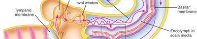
The ear is divided into external, middle, and inner regions, each playing a role in hearing and balance. Sound waves are collected by the external ear, transmitted through the middle ear ossicles, and converted to neural signals in the inner ear.
External Ear: Includes the auricle and ear canal, which direct sound waves to the tympanic membrane.
Middle Ear: Contains the malleus, incus, and stapes, which amplify and transmit vibrations to the oval window.
Inner Ear: The cochlea contains fluid-filled chambers (scala vestibuli, scala media, scala tympani) and the organ of Corti, where hair cells transduce mechanical vibrations into electrical signals sent via the cochlear nerve.
Vestibular System: Includes semicircular canals and otolith organs for balance and spatial orientation.
Muscle Structure and Function
Key Terms and Structures in Muscle Physiology
Muscle tissue is organized into various structural and functional units, each with specific roles in contraction and force generation. Understanding these terms is essential for grasping muscle physiology.
Muscle Fiber: The muscle cell, also known as a myofiber.
Sarcolemma: The plasma membrane of a muscle fiber.
Triad: A T tubule with two lateral sacs of the sarcoplasmic reticulum, crucial for excitation-contraction coupling.
End-Plate Potential (EPP): A graded potential that excites skeletal muscle cells at the neuromuscular junction.
Perimysium: Connective tissue that divides muscles into fascicles.
Lateral Sac: Enlargement of the sarcoplasmic reticulum where calcium is stored.
Endomysium: Thin sheath of connective tissue surrounding individual muscle fibers.
Sarcoplasmic Reticulum (SR): Modified smooth ER that stores calcium.
Epimysium: Connective tissue that surrounds the whole muscle and is continuous with the tendon.
Motor Unit: A somatic motor neuron and all the muscle fibers it innervates.
Myofibril: Rodlike elements containing the contractile machinery of the muscle fiber.
T (Transverse) Tubules: Deep indentations in the sarcolemma that transmit action potentials into the cell.
Sliding Filament Model: The mechanism that drives muscle contraction, involving actin and myosin filaments sliding past each other.
A Band: Region containing both thick and thin filaments; remains the same length during contraction.
M Line: Anchors myosin tails of thick filaments.
Z Line: Anchors actin filaments.
H Zone: Region with only myosin; shortens during contraction.
Actin: Contractile protein making up the thin filament.
Sarcomere: The smallest functional unit of muscle fibers, from Z line to Z line.
DHP Receptors: Voltage sensors in the T tubule membrane.
Ryanodine Receptors: Calcium channels in the SR membrane.
Titin: Structural protein providing support and elasticity.
Tropomyosin: Regulatory protein that blocks myosin-binding sites on actin.
Troponin: Regulatory protein that binds calcium and initiates contraction by moving tropomyosin.
Myosin: Contractile protein making up the thick filament, with heads for cross-bridging.
Comparison of Muscle Types
Muscle tissue is classified into skeletal, smooth (single-unit and multi-unit), and cardiac types, each with distinct structural and functional properties.
Property | Skeletal | Smooth (single unit) | Smooth (multi-unit) | Cardiac |
|---|---|---|---|---|
Striations (sarcomeres) | Yes | No | No | Yes |
Actin and myosin | Yes | Yes | Yes | Yes |
Level of control | Voluntary | Involuntary | Involuntary | Involuntary |
Neural input | Somatic | Autonomic | Autonomic | Autonomic |
Neuroeffector junction | Neuromuscular junction | Diffuse junctions | Diffuse junctions | Intercalated discs |
Hormonal control | No | Yes | Yes | Yes |
Source of calcium | Sarcoplasmic reticulum | SR and extracellular | SR and extracellular | SR and extracellular |
Regulatory protein that binds with calcium | Troponin | Calmodulin | Calmodulin | Troponin |
Gap junctions | No | Yes | No | Yes |
Pacemaker activity | No | Yes | No | Yes |
Myosin ATPase activity | Fast | Slow | Slow | Intermediate |
Cardiac Physiology
Blood Flow Through the Heart
The heart is a muscular organ that pumps blood through the pulmonary and systemic circuits. Blood flow is directed by a series of valves and chambers, ensuring unidirectional movement and efficient oxygenation of tissues.
Right Side: Receives deoxygenated blood from the body and pumps it to the lungs via the pulmonary arteries.
Left Side: Receives oxygenated blood from the lungs and pumps it to the systemic circulation via the aorta.
Valves: Atrioventricular (tricuspid and mitral) and semilunar (pulmonary and aortic) valves prevent backflow.

Cardiac Action Potential
Cardiac muscle cells exhibit a unique action potential with distinct phases, essential for coordinated contraction and relaxation of the heart.
Phase 0 (Depolarization): Rapid influx of Na+ ions causes a sharp rise in membrane potential.
Phase 1 (Initial Repolarization): Brief efflux of K+ ions.
Phase 2 (Plateau): Influx of Ca2+ ions balances K+ efflux, prolonging depolarization.
Phase 3 (Repolarization): K+ efflux restores resting membrane potential.
Phase 4 (Resting Potential): Cell returns to baseline, ready for the next action potential.

