 Back
BackAxial and Appendicular Skeleton, Articulations, and Key Bone Structures – Guided Study Notes
Study Guide - Smart Notes
 Tailored notes based on your materials, expanded with key definitions, examples, and context.
Q1. What are the main regions and features of the axial skeleton?
Background
Topic: Axial Skeleton
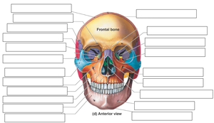
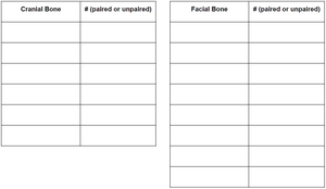
This question focuses on identifying the major bones and features of the axial skeleton, which includes the skull, vertebral column, and thoracic cage.
Key Terms and Concepts:
Axial skeleton: The central part of the skeleton, including the skull, vertebral column, and thoracic cage.
Skull: Protects the brain and forms the structure of the face.
Vertebral column: Supports the body and protects the spinal cord.
Thoracic cage: Protects the heart and lungs.
Step-by-Step Guidance
List the three main regions of the axial skeleton: skull, vertebral column, and thoracic cage.
Identify the major bones in each region (e.g., cranial and facial bones in the skull, cervical/thoracic/lumbar vertebrae, sternum, and ribs).
Consider the functions of each region (protection, support, movement).
Try solving on your own before revealing the answer!
Q2. What are the different types of ribs and how are they classified?
Background
Topic: Thoracic Cage – Ribs
This question tests your understanding of rib classification: true, false, and floating ribs, and their attachment to the sternum.
Key Terms and Concepts:
True ribs: Attach directly to the sternum via costal cartilage.
False ribs: Attach indirectly or not at all to the sternum.
Floating ribs: Do not attach to the sternum.
Step-by-Step Guidance
Recall the total number of ribs in the human body.
Identify which ribs are considered true ribs (typically ribs 1–7).
Determine which ribs are false ribs (typically ribs 8–12), and which of these are floating ribs (usually ribs 11–12).
Try solving on your own before revealing the answer!
Q3. How are the vertebrae organized and what are their unique features?
Background
Topic: Vertebral Column
This question covers the number and types of vertebrae, as well as special features of cervical vertebrae and the sacrum/coccyx.
Key Terms and Concepts:
Cervical, thoracic, lumbar vertebrae: Different regions of the vertebral column.
Atlas (C1): Allows nodding motion.
Axis (C2): Contains the dens, allows rotation.
Transverse foramen: Unique to cervical vertebrae.
Sacrum and coccyx: Fused vertebrae forming the base of the spine.
Step-by-Step Guidance
List the number of vertebrae in each region (cervical, thoracic, lumbar).
Identify the atlas and axis and their functions.
Describe the unique feature of cervical vertebrae (transverse foramen).
Explain the sacrum and coccyx as fused vertebrae.
Try solving on your own before revealing the answer!
Q4. What are the primary and secondary spinal curves?
Background
Topic: Spinal Curvatures
This question asks you to distinguish between the curves present at birth (primary) and those that develop later (secondary).
Key Terms and Concepts:
Primary curves: Thoracic and sacral, present at birth.
Secondary curves: Cervical and lumbar, develop after birth.
Step-by-Step Guidance
Identify which curves are present at birth (primary).
Determine which curves develop as a child grows (secondary).
Try solving on your own before revealing the answer!
Q5. What are the main bones and articulations of the pectoral girdle?
Background
Topic: Appendicular Skeleton – Pectoral Girdle
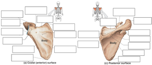
This question focuses on the clavicle and scapula, and their articulations with the sternum and humerus.
Key Terms and Concepts:
Clavicle: Sternal end (articulates with sternum), acromial end (articulates with scapula).
Scapula: Shoulder blade, articulates with humerus.
Step-by-Step Guidance
Identify the two ends of the clavicle and their articulations.
Describe the role of the scapula in the shoulder joint.
Try solving on your own before revealing the answer!
Q6. How do the radius and ulna articulate with the humerus?
Background
Topic: Elbow Joint Anatomy
This question tests your knowledge of the articulating surfaces between the humerus, radius, and ulna.
Key Terms and Concepts:
Capitulum: Articulates with the radius.
Trochlea: Articulates with the ulna.
Head of radius and ulna: Their positions and articulations.
Step-by-Step Guidance
Identify which part of the humerus articulates with the radius (capitulum).
Identify which part of the humerus articulates with the ulna (trochlea).
Describe the location of the head of the radius and ulna.
Try solving on your own before revealing the answer!
Q7. What are the bones of the hand and how are they organized?
Background
Topic: Bones of the Hand
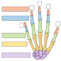
This question asks you to name and number the bones of the hand, including carpals, metacarpals, and phalanges.
Key Terms and Concepts:
Carpals: Wrist bones.
Metacarpals: Palm bones.
Phalanges: Finger bones (proximal, middle, distal).
Step-by-Step Guidance
List the number of carpal, metacarpal, and phalange bones in one hand.
Identify the arrangement of these bones from wrist to fingertips.
Try solving on your own before revealing the answer!
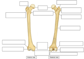
Q8. What are the main features of the femur and patella?
Background
Topic: Lower Limb Bones
This question focuses on the structure and function of the femur and patella, including their anatomical landmarks.
Key Terms and Concepts:
Femur: Thigh bone, largest bone in the body.
Patella: Sesamoid bone, forms the kneecap.
Apex: Bottom of the patella.
Step-by-Step Guidance
Identify the patella as a sesamoid bone.
Locate the apex of the patella.
Describe the main anatomical features of the femur (head, neck, condyles).
Try solving on your own before revealing the answer!
Q9. How are the fibula and tibia positioned, and what are the bones of the pelvis?
Background
Topic: Lower Limb and Pelvic Bones
This question tests your understanding of the medial/lateral orientation of the tibia and fibula, and the names of the pelvic bones (ossa coxae).
Key Terms and Concepts:
Tibia: Medial bone of the lower leg.
Fibula: Lateral bone of the lower leg.
Ossa coxae: Hip bones (ilium, ischium, pubis).
Step-by-Step Guidance
Identify which bone is medial and which is lateral in the lower leg.
List the three bones that make up the os coxae.
Try solving on your own before revealing the answer!
Q10. What are the bones of the foot and their articulations?
Background
Topic: Bones of the Foot
This question asks you to identify the heel bone (calcaneus) and the bone that articulates with the tibia and fibula (talus).
Key Terms and Concepts:
Calcaneus: Heel bone.
Talus: Ankle bone, articulates with tibia and fibula.
Step-by-Step Guidance
Identify the calcaneus as the heel bone.
Identify the talus as the bone that forms the ankle joint with the tibia and fibula.
Try solving on your own before revealing the answer!
Q11. What are the three main types of joints and examples of each?
Background
Topic: Articulations (Joints)
This question covers the classification of joints by movement: diarthrotic (freely movable), amphiarthrotic (slightly movable), and synarthrotic (immovable), with examples.
Key Terms and Concepts:
Diarthrosis: Freely movable joint (e.g., shoulder, knee).
Amphiarthrosis: Slightly movable joint (e.g., pubic symphysis).
Synarthrosis: Immovable joint (e.g., sutures of the skull).
Step-by-Step Guidance
Define each type of joint based on its range of movement.
List at least one example of each type of joint.
Try solving on your own before revealing the answer!
Q12. What are the main ligaments and structures of the knee joint?
Background
Topic: Knee Joint Anatomy
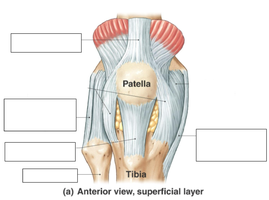
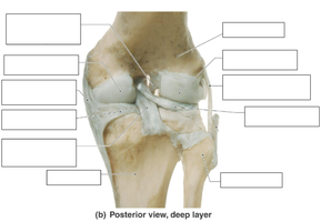
This question asks you to identify the major ligaments and anatomical features of the knee from both anterior and posterior views.
Key Terms and Concepts:
Patella: Kneecap.
Ligaments: ACL, PCL, MCL, LCL, patellar ligament, etc.
Tibia and femur: Bones forming the knee joint.
Step-by-Step Guidance
Study the labeled diagrams of the knee joint (anterior and posterior views).
Identify the patella, tibia, and major ligaments (e.g., anterior cruciate ligament, posterior cruciate ligament, medial/lateral collateral ligaments).
Note the attachments and orientation of each ligament.
Try solving on your own before revealing the answer!
