 Back
BackAxial and Appendicular Skeleton: Identification and Articulation Study Guide
Study Guide - Smart Notes
 Tailored notes based on your materials, expanded with key definitions, examples, and context.
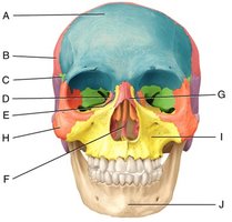
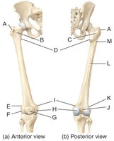
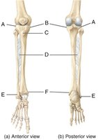
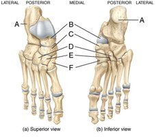
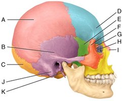
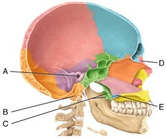
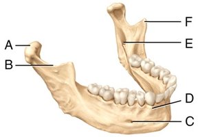
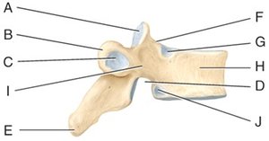
Q1. Identify all labeled structures in the provided images of the skull, femur, tibia/fibula, tarsals, mandible, vertebra, ribs, scapula, and humerus/forearm.
Background
Topic: Human Skeletal System (Axial and Appendicular Skeleton)
This question tests your ability to recognize and name anatomical structures on bones, understand their locations, and relate them to their functions and articulations. This is a foundational skill in anatomy and physiology (ANP) courses, especially for exams covering the skeletal system.
Key Terms and Concepts:
Axial Skeleton: Includes the skull, vertebral column, and thoracic cage (ribs and sternum).
Appendicular Skeleton: Includes the bones of the limbs and girdles (pectoral and pelvic).
Bone Markings: Features such as foramina, condyles, processes, and fossae that serve as attachment points or passageways.
Articulation: Where two bones meet (joint), often marked by specific bone features.
Step-by-Step Guidance
Study each image and note the anatomical orientation (anterior, posterior, lateral, medial, superior, inferior) as labeled. This will help you determine the correct names for each structure.
Recall the major bones and their features for each region. For example, in the skull, identify the frontal, parietal, temporal, occipital, sphenoid, and ethmoid bones, as well as the mandible and maxilla. For the femur, tibia, and fibula, focus on landmarks such as the head, neck, condyles, and malleoli.
Use the labeled letters (A, B, C, etc.) to match each structure to its anatomical name. For example, in the scapula, label the spine, acromion, coracoid process, and glenoid cavity. In the vertebra, identify the body, spinous process, transverse process, and vertebral foramen.
For each bone, consider its articulations with neighboring bones. For example, the humerus articulates with the scapula at the glenoid cavity, and the femur articulates with the acetabulum of the pelvis.
Review the function of each structure. For example, the mandibular condyle allows jaw movement, the olecranon process forms the elbow, and the acetabulum is the socket for the femoral head.
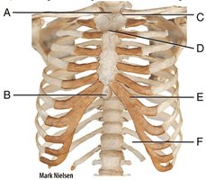
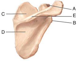
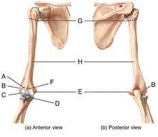
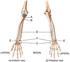
Try solving on your own before revealing the answer!
