 Back
BackAxial and Cranial Bones: Identification and Functions
Study Guide - Smart Notes
 Tailored notes based on your materials, expanded with key definitions, examples, and context.
Q1. Label the major facial and cranial bones on the skull (anterior, posterior, and lateral views).
Background
Topic: Axial Skeleton – Skull Anatomy
This question tests your ability to identify and differentiate between the facial and cranial bones of the skull, as well as understand their anatomical locations and relationships in various views (anterior, posterior, lateral).
Key Terms and Concepts:
Facial Bones: Bones that form the structure of the face (e.g., nasal, maxillae, zygomatic, mandible).
Cranial Bones: Bones that form the cranium, protecting the brain (e.g., frontal, parietal, temporal, occipital, sphenoid, ethmoid).
Sutures: Immovable joints connecting the bones of the skull.
Landmarks: Specific features such as processes, foramina, and fossae that serve as attachment points or passageways.
Step-by-Step Guidance
Start by distinguishing between facial bones (which form the face and jaw) and cranial bones (which encase the brain). Review the list of paired and single bones in each category.
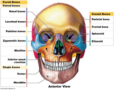
On the anterior view, identify the paired facial bones (nasal, lacrimal, palatine, zygomatic, maxillae, inferior nasal conchae) and single bones (vomer, mandible). For cranial bones, locate the frontal, parietal, sphenoid, and ethmoid bones.
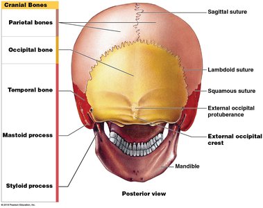
On the posterior view, focus on the cranial bones: parietal, occipital, and temporal bones. Identify the major sutures (sagittal, lambdoid, squamous) and bony landmarks such as the external occipital protuberance and crest.
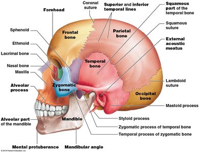
On the lateral view, identify the relationships between the cranial bones (frontal, parietal, temporal, occipital, sphenoid, ethmoid) and facial bones (zygomatic, maxilla, mandible, nasal, lacrimal). Note the location of important processes and sutures.
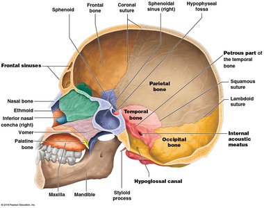
Review the internal and inferior views to see how the bones articulate and where foramina (holes) are located for nerves and blood vessels. Pay attention to the sphenoid, ethmoid, and occipital bones in these views.
Try solving on your own before revealing the answer!
Final Answer: Key Skull Bones and Sutures Labeled
On the anterior view, the paired facial bones are the nasal, lacrimal, palatine, zygomatic, maxillae, and inferior nasal conchae. The single facial bones are the vomer and mandible. The cranial bones visible are the frontal, parietal, sphenoid, and ethmoid.
On the posterior view, the cranial bones are the parietal, occipital, and temporal bones, with the sagittal, lambdoid, and squamous sutures connecting them.
On the lateral view, you can see the frontal, parietal, temporal, occipital, sphenoid, and ethmoid cranial bones, as well as the zygomatic, maxilla, mandible, nasal, and lacrimal facial bones. Key features include the mastoid and styloid processes, zygomatic arch, and alveolar processes.
