 Back
BackAxial Skeleton and Cranial Bones: Structure and Key Features
Study Guide - Smart Notes
 Tailored notes based on your materials, expanded with key definitions, examples, and context.
Lab 3: Axial Skeleton
Overview of the Skeleton
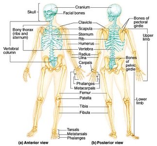
The human skeleton is a structural framework composed of bones, cartilage, joints, and ligaments. It is divided into two main divisions: the axial skeleton and the appendicular skeleton. The axial skeleton consists of 80 bones, while the appendicular skeleton contains 126 bones.
Axial Skeleton: Includes the skull, vertebral column, and bony thorax.
Appendicular Skeleton: Comprises the limbs and girdles that attach them to the axial skeleton.
The Axial Skeleton
Main Components
The axial skeleton forms the central axis of the body and is essential for protection and support. It is made up of the skull, vertebral column, and bony thorax.
Skull: Protects the brain and forms the structure of the face.
Vertebral Column: Supports the body and protects the spinal cord.
Bony Thorax: Includes the ribs and sternum, protecting the heart and lungs.
Bone Markings
Types and Functions
Bones display various markings that serve as sites for muscle and ligament attachment, form joints, or allow passage for nerves and blood vessels.
Projections: Provide attachment points for muscles and ligaments.
Joint-forming Projections: Help form articulations between bones.
Depressions and Openings: Allow passage of nerves and blood vessels.
Key Anatomical Terms
Definitions
Understanding anatomical terminology is crucial for identifying bone features and articulations.
Foramen: A hole or opening in a bone.
Fossae: Basin-like depressions in bone.
Suture: An immovable fibrous joint between bones of the skull.
Articulation: A joint between bones.
Articulate: To form a joint.
Ramus: A bony projection.
Condyle: A smooth prominence on a bone where it forms a joint with another bone.
Concha: A bone curled like a sea shell.
Cranial Bones
Structure and Function
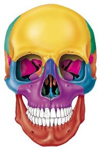
The cranial bones form the protective case around the brain and provide attachment points for head and neck muscles. There are eight cranial bones: frontal, parietal, temporal, sphenoid, ethmoid, and occipital.
Frontal Bone: Forms the forehead and the upper part of the eye sockets.
Parietal Bones: Form the sides and roof of the cranial cavity.
Temporal Bones: Located at the sides and base of the skull.
Sphenoid Bone: Forms part of the base of the skull and the sides of the eye sockets.
Ethmoid Bone: Located between the eyes, forms part of the nasal cavity.
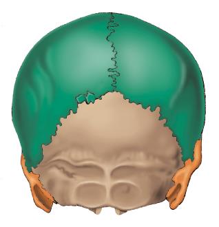
Occipital Bone: Forms the back and base of the skull.

Sutures
Types and Locations
Sutures are immovable joints found only between the bones of the skull. They are important for the stability and protection of the brain.
Coronal Suture: Between the frontal and parietal bones.
Squamous Suture: Between the parietal and temporal bones.
Lambdoid Suture: Between the parietal and occipital bones.
Sagittal Suture: Between the two parietal bones.
Facial Bones
Structure and Function
The facial bones form the structure of the face, support the teeth, and provide attachment points for facial muscles. Key facial bones include the nasal bone, mandible, zygomatic bone, maxilla, vomer, lacrimal, and inferior nasal concha.
Nasal Bone: Forms the bridge of the nose.
Mandible: The lower jawbone, the only movable bone of the skull.
Zygomatic Bone: Forms the cheekbones.
Maxilla: Forms the upper jaw and part of the hard palate.
Vomer: Forms part of the nasal septum.
Lacrimal Bone: Small bone forming part of the eye socket.
Inferior Nasal Concha: Curved bone in the nasal cavity.
Summary Table: Major Cranial and Facial Bones
Bone | Location | Function |
|---|---|---|
Frontal | Forehead, upper eye sockets | Protection, muscle attachment |
Parietal | Sides and roof of skull | Protection |
Temporal | Sides and base of skull | Protection, muscle attachment |
Sphenoid | Base of skull, sides of eye sockets | Support, muscle attachment |
Ethmoid | Between eyes, nasal cavity | Support, olfactory function |
Occipital | Back and base of skull | Protection, muscle attachment |
Mandible | Lower jaw | Chewing, speech |
Maxilla | Upper jaw | Chewing, support teeth |
Zygomatic | Cheek | Facial structure |
Nasal | Bridge of nose | Support nose |
