 Back
BackAxial Skeleton: Bones and Features for Identification
Study Guide - Smart Notes
 Tailored notes based on your materials, expanded with key definitions, examples, and context.
Axial Skeleton Overview
The axial skeleton forms the central axis of the human body and includes the skull, vertebral column, and thoracic cage. It provides support, protection for vital organs, and attachment points for muscles.
Skull
Cranial Bones
The cranial bones protect the brain and form the structure of the head. Key bones and features include:
Frontal (1): Contains the frontal sinuses.
Parietal (2): Marked by temporal lines.
Occipital (1): Features the occipital condyles (articulate with the atlas) and the foramen magnum (passage for the spinal cord).
Temporal (2): Includes petrous and squamous parts, external acoustic meatus, mastoid process, zygomatic process, and mandibular fossa.
Sphenoid (1): Contains greater and lesser wings, sella turcica (houses pituitary gland), optic canals, and sphenoid sinus.
Ethmoid: Features cribriform plate, perpendicular plate (forms part of nasal septum), and superior and middle nasal conchae.
Facial Bones
Zygomatic (2): Forms the cheekbones; temporal process is part of the zygomatic arch.
Lacrimal (2): Contains the lacrimal fossa for tear ducts.
Nasal (2): Forms the bridge of the nose.
Inferior nasal conchae (2): Increases surface area in the nasal cavity.
Maxillary (2): Contains maxillary sinuses and alveolar process (holds teeth).
Vomer (1): Forms the lower part of the nasal septum.
Palatine (2, fused): Forms the posterior part of the hard palate.
Mandible (1): Lower jaw; features alveolar processes, body, angle, rami, coronoid processes, and condylar processes (articulate with temporal bone at TMJ).
Sutures and Fontanelles
Sutures: Immovable joints between skull bones. Major sutures include coronal, sagittal, squamous, and lambdoid.
Fontanelles: Soft spots in the fetal skull that allow for growth and flexibility during birth.
Hyoid Bone
The hyoid bone is unique as it does not articulate with any other bone. Located in the throat, it supports the larynx and is anchored by neck muscles.
Vertebral Column
The vertebral column supports the body, protects the spinal cord, and provides attachment points for ribs and muscles. It consists of five regions:
Cervical (7): Neck region
Thoracic (12): Upper back, articulates with ribs
Lumbar (5): Lower back, supports most body weight
Sacral (5, fused): Forms the sacrum
Coccygeal (3-5, fused): Forms the coccyx (tailbone)
Intervertebral discs separate vertebrae and act as shock absorbers.
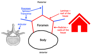
Typical Vertebra Structure
Body: Weight-bearing portion
Vertebral arch: Formed by pedicles and laminae
Vertebral foramen: Passage for the spinal cord
Processes: Transverse, spinous, superior & inferior articular processes
Cervical Vertebrae (C1–C7)
Typical cervical vertebrae: Small, with transverse foramina for vertebral arteries, bifid spinous processes, and short transverse processes.
Atlas (C1): Lacks a body and spinous process; has anterior and posterior arches; articulates with occipital condyles for nodding motion.
Axis (C2): Has a dens (odontoid process) that allows rotation of the head.
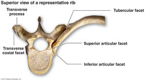
Thoracic Vertebrae (T1–T12)
Medium-sized, heart-shaped body with superior and inferior costal facets for rib articulation.
Long, downward-pointing spinous processes.
Transverse costal facets on transverse processes for rib tubercles.
Lumbar Vertebrae (L1–L5)
Large, robust bodies for weight bearing.
Short, thick spinous processes and long, slender transverse processes.
Sacrum and Coccyx
Sacrum: Five fused vertebrae; features median sacral crest and sacral hiatus.
Coccyx: Three to five fused segments; serves as an attachment for ligaments and muscles.
Thoracic Skeleton
Ribs
There are 12 pairs of ribs, each articulating with thoracic vertebrae. Key features include:
Head: Has articular facets for vertebral bodies.
Neck: Connects head to shaft.
Tubercle: Contains tubercular facet for articulation with transverse costal facet of vertebra.
Shaft: Main body of the rib.
Costal cartilages: Connect ribs to sternum (ribs I–VII) or to cartilage above (ribs VIII–X); ribs XI and XII are floating ribs with no costal cartilage.
Sternum
Manubrium: Superior portion
Body: Central portion
Xiphoid process: Inferior tip
Summary Table: Vertebral Regions and Key Features
Region | Number of Vertebrae | Key Features |
|---|---|---|
Cervical | 7 | Transverse foramina, bifid spinous process, atlas & axis |
Thoracic | 12 | Costal facets for ribs, long spinous process |
Lumbar | 5 | Large body, short spinous process |
Sacral | 5 (fused) | Median sacral crest, sacral hiatus |
Coccygeal | 3–5 (fused) | Small, fused segments |
Additional info:
Fontanelles allow for brain growth and skull flexibility in infants.
The temporomandibular joint (TMJ) is formed by the articulation of the mandibular condyle with the mandibular fossa of the temporal bone.
Intervertebral discs are composed of an outer annulus fibrosus and an inner nucleus pulposus, providing cushioning between vertebrae.
