 Back
BackL2: Axial - Skull
Study Guide - Smart Notes
 Tailored notes based on your materials, expanded with key definitions, examples, and context.
The Axial Skeleton
Overview
The axial skeleton forms the central axis of the human body, consisting of the skull, vertebral column, and thoracic cage. It serves to protect vital organs such as the brain, spinal cord, heart, and lungs, and provides attachment sites for muscles involved in movement and posture.
Total bones: 80 in adults
Main divisions: Skull, vertebral column, thoracic cage
Functions: Protection, support, muscle attachment
Skull
Cranium (Cranial Vault, Braincase)
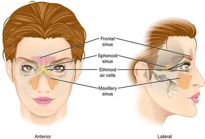
The cranium encloses and protects the brain and provides attachment points for muscles involved in chewing and head movement. It contains air-filled sinuses that reduce weight and contribute to resonance.
Single cranial bones: Frontal, occipital, sphenoid, ethmoid
Paired cranial bones: Parietal, temporal
Sinuses: Found in frontal, sphenoid, ethmoid, and maxillary bones
Sutures: Coronal, sagittal, lambdoid, squamous
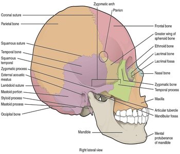
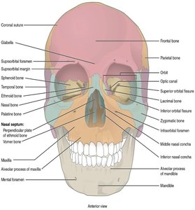
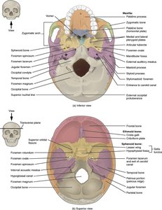
Key Cranial Bone Features
Frontal bone: Forms forehead, roof of nasal cavity, roof of orbits; contains frontal sinuses
Occipital bone: Forms back and base of cranium; features foramen magnum and occipital condyles
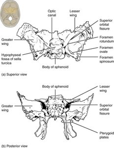
Sphenoid bone: Forms base and sides of cranium; houses sella turcica (pituitary gland); contains two sinuses
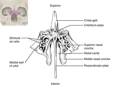
Ethmoid bone: Forms part of nasal cavity roof, cranial floor, orbital walls; features crista galli, cribriform plate, perpendicular plate, and sinuses
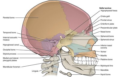
Facial Bones
The facial bones protect sensory organs and provide attachment for muscles of mastication, facial expression, and eye movement. There are thirteen immovable and one movable bone (mandible).
Single facial bones: Mandible (lower jaw), vomer (lower nasal septum)
Paired facial bones: Maxillae (upper jaw), palatine, zygomatic (cheek bones), lacrimal, nasal, inferior nasal conchae
Key Facial Bone Features
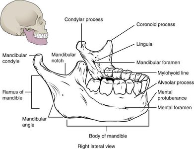
Mandible: Only movable bone; features include body, ramus, mandibular notch, condyle, coronoid process, mental foramen, alveolar processes
Maxillae: Form upper jaw, roof of mouth, house largest sinuses, alveolar processes
Zygomatic bones: Form cheek prominences, floor/lateral walls of orbits
Palatine bones: Form part of hard palate
Vertebral Column
General Structure
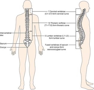
The vertebral column consists of 26 vertebrae in adults, separated by intervertebral discs. It protects the spinal cord and supports the body.
Regions: Cervical (7), thoracic (12), lumbar (5), sacrum (5 fused), coccyx (4 fused)
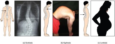
Curvatures: Primary (thoracic, sacral), secondary (cervical, lumbar)
Abnormalities: Kyphosis (hunchback), lordosis (swayback), scoliosis (lateral curve)
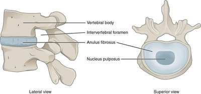
Structure of a Typical Vertebra
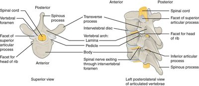
Centrum (body): Large ventral portion
Vertebral arch: Laminae (dorsal arch), pedicles (lateral portions), vertebral foramen
Processes: Spinous, transverse, superior/inferior articular
Regional Differences
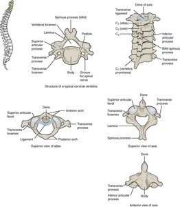
Cervical vertebrae: Smallest, bifid spinous processes (C2-6), transverse foramina, atlas (C1), axis (C2 with dens)
Thoracic vertebrae: Long, pointed spinous processes, articular surfaces for ribs
Lumbar vertebrae: Largest, horizontal spinous process, large centrum
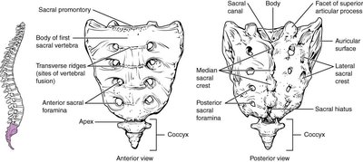
Sacrum: 5 fused vertebrae, dorsal/ventral foramina, sacral promontory, sacroiliac joint
Coccyx: 4 fused vertebrae, shock absorber
Thoracic Cage
Structure and Function
The thoracic cage supports the pectoral girdle and arms, protects thoracic and upper abdominal organs, and aids in breathing.
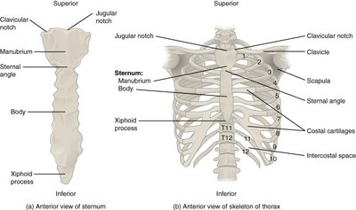
Ribs: 12 pairs attached dorsally to thoracic vertebrae
True ribs: First 7 pairs, attach directly to sternum
False ribs: Pairs 8-12, do not attach directly to sternum; last 2 pairs are floating ribs
Sternum: Manubrium (superior), body (middle), xiphoid process (inferior)
Identification and Review
Skull and Thoracic Cage Items for Identification
Skull: Condylar process, palatine bone, coronal suture, occipital bone, coronoid process, parietal bone, cribriform plate, occipital condyle, crista galli, olfactory foramina, ethmoid bone, perpendicular plate, external auditory meatus, sagittal suture, foramen magnum, sella turcica, frontal bone, sphenoid bone, hyoid bone, squamous suture, incisive foramen, styloid process, lacrimal bone, supraorbital foramen, lambdoid suture, temporal bone, mandible, vomer bone, mastoid process, zygomatic bone, maxillae, zygomatic process, mental foramen, nasal bone, nasal conchae (superior/middle/inferior)
Thoracic Cage and Spine: Atlas, vertebral column, axis, suprasternal notch, body, sternum, cervical vertebrae, thoracic vertebrae, coccyx, transverse process, intervertebral discs, vertebral ribs (floating), lamina, vertebrochondral ribs (false), lumbar vertebrae, transverse foramina, manubrium (body), vertebral foramen, pedicle, vertebrosternal ribs (true), sacrum, xiphoid process, spinous process
Lab Review and Problem Solving
Sample Questions and Explanations
External acoustic meatus location: Temporal bone
Spinous process projection: Thoracic vertebra
Teeth association: Maxilla and mandible
Hard palate formation: Maxilla and palatine bones
Kyphosis: Primary thoracic curvature abnormality
Pituitary gland location: Sella turcica of sphenoid bone
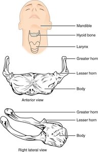
Skull exception: Hyoid bone is not part of the skull
Lambdoid suture: Joins parietal and occipital bones
Cervical vertebrae: 7 in cervical region
Spinal cord exit: Foramen magnum
Problem Solving Example
Joe's lumbar scoliosis after polio:
Accept: Paralysis during polio resulted in unequal muscle pull on the spine, leading to abnormal curvature (scoliosis).
Reject: Osteoporosis/osteomalacia, kyphosis, obesity, and unrelated post-growth causes are not supported by the clinical history.
Summary Table: Cranial vs. Facial Bones
Bone | Type |
|---|---|
Ethmoid | Cranial |
Frontal | Cranial |
Inferior nasal concha | Facial |
Lacrimal | Facial |
Mandible | Facial |
Maxilla | Facial |
Nasal | Facial |
Occipital | Cranial |
Palatine | Facial |
Parietal | Cranial |
Sphenoid | Cranial |
Temporal | Cranial |
Vomer | Facial |
Zygomatic | Facial |
Additional info:
Sinuses are air-filled spaces that lighten the skull and enhance voice resonance.
Intervertebral discs consist of anulus fibrosus (outer ring) and nucleus pulposus (inner core).
Abnormal spinal curvatures can result from muscle imbalance, congenital defects, or trauma.
