 Back
BackBIO 163: Skeletal, Muscular, and Nervous System Study Guide
Study Guide - Smart Notes
 Tailored notes based on your materials, expanded with key definitions, examples, and context.
Skeletal and Muscular Systems
Types of Muscle Tissue
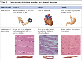
Muscle tissue is classified into three main types: skeletal, cardiac, and smooth. Each type has distinct structural and functional characteristics that suit its role in the body.
Skeletal Muscle: Attached to bones or, for some facial muscles, to skin. Responsible for voluntary movements.
Cardiac Muscle: Found only in the walls of the heart. Responsible for pumping blood throughout the body.
Smooth Muscle: Located in the walls of hollow organs (other than the heart), such as the stomach and intestines. Involved in involuntary movements like peristalsis.
Characteristic | Skeletal | Cardiac | Smooth |
|---|---|---|---|
Body location | Attached to bones or skin | Walls of the heart | Walls of hollow organs |
Cell shape & appearance | Single, very long, cylindrical, multinucleate cells with obvious striations | Branching chains of cells; uninucleate, striations; intercalated discs | Single, fusiform, uninucleate; no striations |
Nervous System
Major Brain Regions and Their Functions
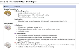
The brain is divided into several major regions, each with specialized functions essential for sensory processing, motor control, and regulation of homeostasis.
Cerebral Hemispheres: Involved in sensory input localization, voluntary muscle activity, and intellectual processing.
Diencephalon: Includes the thalamus (sensory relay), hypothalamus (autonomic and endocrine regulation), and limbic system (emotions and memory).
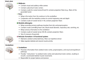
Brain Stem: Contains centers for visual/auditory reflexes, respiratory and cardiovascular control, and relays information between brain regions.
Cerebellum: Coordinates voluntary movements and balance.
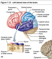
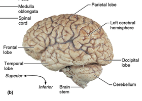
Brain Anatomy: Lobes, Sulci, and Fissures
The brain's surface is marked by elevated ridges (gyri), shallow grooves (sulci), and deeper grooves (fissures). These features divide the brain into lobes, each associated with specific functions.
Frontal Lobe: Voluntary movement, planning, reasoning, and speech production (Broca's area).
Parietal Lobe: Sensory perception and integration.
Temporal Lobe: Auditory processing and memory (Wernicke's area for language comprehension).
Occipital Lobe: Visual processing.
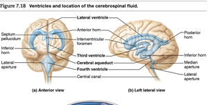
Ventricles and Cerebrospinal Fluid (CSF)
The brain contains interconnected cavities called ventricles, which are filled with cerebrospinal fluid (CSF). CSF cushions the brain, removes waste, and circulates nutrients.
Lateral Ventricles: Largest ventricles, located in each cerebral hemisphere.
Third Ventricle: Located in the diencephalon.
Fourth Ventricle: Located between the brainstem and cerebellum.
CSF Circulation: CSF is produced by the choroid plexuses, flows through the ventricles, enters the subarachnoid space, and is absorbed into the venous blood.
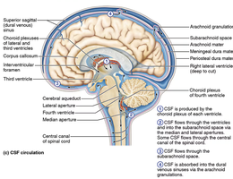
Additional info:
CSF Function: Protects the brain and spinal cord from trauma, supplies nutrients, and removes waste products.
Clinical Application: Blockage of CSF flow can lead to hydrocephalus, a condition characterized by increased intracranial pressure.
