 Back
BackBIO 163: Skeletal, Muscular, and Nervous System Study Guide
Study Guide - Smart Notes
 Tailored notes based on your materials, expanded with key definitions, examples, and context.
Skeletal and Muscular Systems
Types of Muscle Tissue
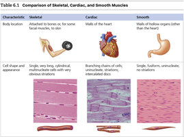
Muscle tissue is classified into three main types: skeletal, cardiac, and smooth. Each type has distinct structural and functional characteristics that are essential for various bodily functions.
Skeletal Muscle: Attached to bones or, for some facial muscles, to skin. Cells are long, cylindrical, multinucleate, and show obvious striations.
Cardiac Muscle: Found only in the walls of the heart. Cells are branching chains, striated, and interconnected by intercalated discs.
Smooth Muscle: Located in the walls of hollow organs (other than the heart). Cells are single, fusiform, uninucleate, and lack striations.
Example: Skeletal muscle enables voluntary movement, cardiac muscle contracts to pump blood, and smooth muscle controls involuntary movements in organs like the stomach.
Neuromuscular Junction
The neuromuscular junction is the region where a motor neuron comes into close contact with a skeletal muscle cell. It is essential for transmitting nerve impulses that trigger muscle contraction.
Key Point: Neurotransmitters released from the axon terminal stimulate the muscle cell, leading to contraction.
Example: Acetylcholine is the primary neurotransmitter at the neuromuscular junction.
Movements at Joints
Joints allow for various types of movement, which are classified based on the direction and nature of the motion.
Flexion: Decreases the angle between two bones (e.g., bending the knee).
Extension: Increases the angle between two bones (e.g., straightening the elbow).
Rotation: Movement of a bone around its longitudinal axis (e.g., shaking head "no").
Abduction: Moving a limb away from the midline.
Adduction: Moving a limb toward the midline.
Circumduction: Combination of flexion, extension, abduction, and adduction.
Special Movements: Dorsiflexion, plantar flexion, inversion, eversion, supination, pronation, opposition.
Example: Flexion at the elbow brings the forearm closer to the upper arm.
Origins and Insertions of Muscles
Muscles are named based on their origin and insertion points, as well as the number of origins.
Number of Origins: Biceps (two), triceps (three), quadriceps (four).
Location: Sternocleidomastoid originates from the sternum and clavicle, inserts on the mastoid process.
Interchangeable Origins/Insertions: Some muscles, like the rectus femoris, can have interchangeable origins and insertions depending on the action.
Nervous System
Functions of the Nervous System
The nervous system has three overlapping functions: sensory input, integration, and motor output.
Sensory Input: Monitors changes (stimuli) inside and outside the body.
Integration: Processes and interprets sensory input to decide on a response.
Motor Output: Activates muscles or glands to cause a response.
Organization of the Nervous System
The nervous system is organized into the central nervous system (CNS) and peripheral nervous system (PNS).
CNS: Brain and spinal cord; command centers for interpreting sensory information and issuing instructions.
PNS: Cranial and spinal nerves; communication lines linking the body to the CNS.
Functional Classification of the PNS
The PNS is divided into sensory (afferent) and motor (efferent) divisions.
Sensory (Afferent): Carries impulses to the CNS from sensory receptors.
Motor (Efferent): Carries impulses from the CNS to effector organs.
Motor Division: Somatic vs. Autonomic
The motor division is further divided into somatic (voluntary) and autonomic (involuntary) systems.
Somatic: Controls voluntary movements of skeletal muscles.
Autonomic: Regulates involuntary activities of smooth muscle, cardiac muscle, and glands.
Neuroglia and Myelin Sheath
Neuroglia are supporting cells in the CNS and PNS. Myelin sheath is a protective layer that insulates axons and speeds up nerve impulse transmission.
CNS: Oligodendrocytes form myelin sheaths.
PNS: Schwann cells form myelin sheaths.
Function: Myelinated fibers conduct impulses faster via saltatory conduction.
Nodes of Ranvier
Nodes of Ranvier are gaps in the myelin sheath that facilitate rapid impulse transmission.
Importance: Allow impulses to jump from node to node, increasing speed.
Structure and Function of Neurons
Neurons have specialized structures for transmitting electrical signals.
Dendrites: Carry signals to the cell body.
Axons: Carry signals away from the cell body.
Axon Hillock: Origin of the axon.
Axon Terminals: Branches that form synapses with other cells.
Neurotransmitters: Chemicals that transmit signals across synapses.
Synaptic Cleft: Space between neurons at a synapse.
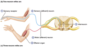
Reflex Arcs
Reflex arcs are neural pathways that mediate rapid, predictable, and involuntary responses to stimuli. They consist of five elements: receptor, sensory neuron, integration center, motor neuron, and effector.
Order: 1) Receptor, 2) Sensory neuron, 3) Integration center, 4) Motor neuron, 5) Effector
Types: Stretch reflex (single synapse), withdrawal reflex (interneurons involved)
Brain Anatomy and Function
Major Brain Regions and Their Functions
The brain is divided into several regions, each with specialized functions.
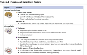
Cerebral Hemispheres: Localize and interpret sensory input, control voluntary muscle activity, and process intellect and emotion.
Diencephalon: Includes thalamus (relay station), hypothalamus (autonomic control, homeostasis), and limbic system (emotion, memory).
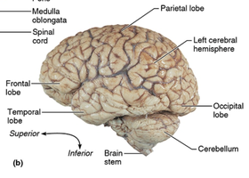
Brain Stem: Midbrain (visual/auditory reflexes), pons (breathing control), medulla oblongata (cardiovascular, respiratory centers), reticular formation (consciousness).
Cerebellum: Coordinates skeletal muscle activity and balance.

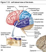
Lobes, Sulci, and Fissures of the Cerebral Cortex
The cerebral cortex is divided into lobes by sulci (shallow grooves) and fissures (deep grooves). Each lobe is associated with specific functions.
Frontal Lobe: Motor control, problem-solving, speech.
Parietal Lobe: Sensory processing.
Temporal Lobe: Auditory processing, memory.
Occipital Lobe: Visual processing.
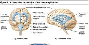
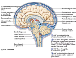
Ventricles and Cerebrospinal Fluid (CSF)
The brain contains ventricles filled with cerebrospinal fluid, which cushions and protects neural tissue.
Ventricles: Lateral, third, and fourth ventricles are interconnected and circulate CSF.
CSF Function: Provides nutrients, removes waste, and maintains pressure.
Additional info:
Tables and diagrams included above are recreated from textbook-style figures to aid understanding of muscle types, reflex arcs, brain regions, and CSF flow.
Key terms and concepts are expanded for clarity and exam preparation.
