 Back
BackBIO 201 Lab Final Study Guide: Muscles, Nervous System, and Special Senses
Study Guide - Smart Notes
 Tailored notes based on your materials, expanded with key definitions, examples, and context.
Chapter 9: Muscles and Muscle Tissue
Muscle Contraction and Types
Muscle contraction is the process by which muscle fibers generate tension and produce movement or maintain posture. The relationship between muscle tension and the load determines the type of contraction.
Muscle Tension: The force exerted by a contracting muscle on an object.
Load: The opposing force exerted by the object on the muscle.
Isotonic Contraction: Muscle changes length as tension rises. Two types:
Concentric Contraction: Muscle tension exceeds load; muscle shortens and moves the object.
Eccentric Contraction: Load exceeds muscle tension; muscle lengthens during contraction.
Isometric Contraction: Muscle tension increases but does not overcome the load; muscle length remains constant.
Muscle Fatigue and Recruitment
Muscle Fatigue: Condition where a muscle loses its ability to generate force.
Threshold Stimulus: Minimum stimulus required to initiate a muscle fiber contraction.
Motor Unit Recruitment: Activation of increasing numbers of motor units to increase muscle force.
Motor Unit: A motor nerve fiber and all the muscle fibers it controls.
Muscle Flexibility: The range of motion at a joint.
Muscle Structure: Actin and Myosin
Actin: Thin filament composed of monomeric subunits with myosin-binding sites; regulated by troponin and tropomyosin.
Myosin: Thick filament with heads that pivot and bend; hydrolyzes ATP to form crossbridges with actin.
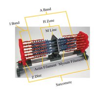
Additional info: The sarcomere is the functional unit of muscle contraction, defined by the region between two Z discs. The arrangement of actin (thin) and myosin (thick) filaments creates the striated appearance of skeletal muscle.
Chapter 10: The Muscular System
Muscle Identification and Anatomy
Understanding the anatomy of skeletal muscles includes identifying major muscle groups and their attachments.
Head and Neck Muscles
Abdominal Muscles
Posterior Back Muscles
Posterior Leg Muscles
Key muscles to know (including origin, action, and insertion):
Rectus Abdominis
Pectoralis Major
Deltoid
Serratus Anterior
Trapezius
Biceps Brachii
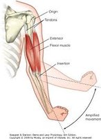
Additional info: The origin is the fixed attachment, while the insertion moves with contraction. Muscles work in pairs: flexors decrease the angle at a joint, extensors increase it.
Muscle Microanatomy
Skeletal muscle is organized into bundles of fibers, each surrounded by connective tissue layers.
Epimysium: Surrounds the entire muscle.
Perimysium: Surrounds bundles of muscle fibers (fascicles).
Endomysium: Surrounds individual muscle fibers.
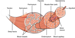
Additional info: Each muscle fiber contains myofibrils, which are composed of repeating sarcomeres.
Chapter 11: Fundamentals of the Nervous System and Nervous Tissue
Chicken Dissection Lab
Dissection labs help identify anatomical structures and understand their functions. Review all structures identified in the chicken dissection and complete the worksheet provided.
Chapter 12: The Central Nervous System
Spinal Cord and Brain Anatomy
Be able to identify the following structures:
Spinal Cord Cross Section: Recognize gray matter, white matter, dorsal and ventral horns.
Sagittal Brain Section: Identify major regions such as the cerebrum, cerebellum, brainstem, and ventricles.
Cerebral Cortex Areas: Match areas (e.g., Wernicke’s area, Broca’s area) with their associated symptoms.
Additional info: Wernicke’s area is involved in language comprehension; Broca’s area is involved in speech production.
Chapter 13: The Peripheral Nervous System and Reflex Activity
Cranial Nerves
Be able to identify all cranial nerves and classify them as sensory, motor, or both.
Cranial Nerve | Function | Type |
|---|---|---|
Olfactory (I) | Smell | Sensory |
Optic (II) | Vision | Sensory |
Oculomotor (III) | Eye movement | Motor |
Trochlear (IV) | Eye movement | Motor |
Trigeminal (V) | Facial sensation, chewing | Both |
Abducens (VI) | Eye movement | Motor |
Facial (VII) | Facial expression, taste | Both |
Vestibulocochlear (VIII) | Hearing, balance | Sensory |
Glossopharyngeal (IX) | Taste, swallowing | Both |
Vagus (X) | Viscera, speech | Both |
Accessory (XI) | Neck muscles | Motor |
Hypoglossal (XII) | Tongue movement | Motor |
Special Senses: Eye and Brain Dissections
Cow Eye Dissection
Know the structures of the cow eye, including the cornea, lens, retina, optic nerve, and associated layers.
Sheep Brain Dissection
Identify the major structures of the sheep brain and the cranial nerves.
