 Back
BackBIO 2121 Exam 6 Review – Anatomy & Physiology Study Guidance
Study Guide - Smart Notes
 Tailored notes based on your materials, expanded with key definitions, examples, and context.
Q18. Identify A-E in the eye diagram below:
Background
Topic: Anatomy of the Eye
This question tests your knowledge of the major anatomical structures of the eye, which are essential for understanding vision and the function of each part in the visual process.
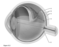
Key Terms:
Cornea: The transparent front part of the eye that covers the iris and pupil.
Lens: Focuses light onto the retina.
Retina: Contains photoreceptor cells (rods and cones) that detect light.
Optic Nerve: Transmits visual information from the retina to the brain.
Iris: The colored part of the eye that controls the size of the pupil.
Step-by-Step Guidance
Observe the diagram and note the location of each labeled structure (A-E).
Recall the anatomical position of the cornea (front), lens (behind cornea), retina (lining the back), optic nerve (exiting the back), and iris (surrounding the pupil).
Match each label (A-E) to the correct anatomical structure based on its position in the diagram.
Write down your answers for each label before checking the final answer.
Try solving on your own before revealing the answer!
Final Answer:
A: Cornea
B: Iris
C: Lens
D: Retina
E: Optic Nerve
These are the main anatomical structures of the eye as labeled in the diagram. Knowing their locations and functions is crucial for understanding how vision works.
Q19. Identify A-E in the eye diagram below:
Background
Topic: Anatomy of the Eye (Alternate View)
This question also tests your ability to identify key structures of the eye, possibly from a slightly different perspective or with different labeling.
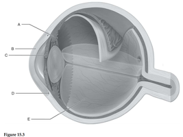
Key Terms:
Pupil: The opening in the center of the iris that allows light to enter the eye.
Lens: Focuses incoming light onto the retina.
Retina: The sensory membrane that lines the back of the eye.
Vitreous Humor: The clear gel that fills the space between the lens and the retina.
Optic Nerve: Carries visual signals to the brain.
Step-by-Step Guidance
Examine the diagram and locate each labeled part (A-E).
Use your knowledge of eye anatomy to identify each structure based on its position and appearance.
Remember the order of structures from front (cornea/pupil) to back (retina/optic nerve).
Assign the correct anatomical name to each label.
Try solving on your own before revealing the answer!
Final Answer:
A: Sclera
B: Choroid
C: Retina
D: Vitreous Humor
E: Optic Nerve
These structures are essential for the function and support of the eye. The sclera is the white outer layer, the choroid provides blood supply, and the vitreous humor maintains eye shape.
Q20. Identify A-E in the ear diagram below:
Background
Topic: Anatomy of the Ear
This question assesses your understanding of the main anatomical parts of the ear, which are involved in hearing and balance.
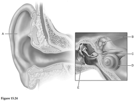
Key Terms:
Pinna (Auricle): The external part of the ear that collects sound waves.
Tympanic Membrane (Eardrum): Vibrates in response to sound waves.
Cochlea: Spiral-shaped organ responsible for hearing.
Pharyngotympanic (Eustachian) Tube: Equalizes pressure between the middle ear and the atmosphere.
Ossicles: Small bones (malleus, incus, stapes) that transmit sound vibrations.
Step-by-Step Guidance
Carefully observe the diagram and the location of each label (A-E).
Recall the anatomical layout of the ear: outer ear (pinna), middle ear (ossicles, tympanic membrane), and inner ear (cochlea, vestibular apparatus).
Match each label to the correct structure based on its position and function.
Write down your answers for each label before checking the final answer.
Try solving on your own before revealing the answer!
Final Answer:
A: Pinna (Auricle)
B: Tympanic Membrane (Eardrum)
C: Cochlea
D: Pharyngotympanic (Eustachian) Tube
E: Ossicles
These are the main structures involved in hearing and balance. The pinna collects sound, the tympanic membrane vibrates, the ossicles transmit vibrations, the cochlea processes sound, and the Eustachian tube equalizes pressure.
Q21. Identify A-E in the endocrine glands diagram below:
Background
Topic: Endocrine System Anatomy
This question tests your ability to identify the major endocrine glands in the human body, which secrete hormones that regulate various physiological processes.
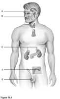
Key Terms:
Pituitary Gland: The "master gland" that controls other endocrine glands.
Thyroid Gland: Regulates metabolism.
Pancreas: Regulates blood glucose levels.
Adrenal Glands: Produce hormones like cortisol and adrenaline.
Gonads (Testes/Ovaries): Produce sex hormones.
Step-by-Step Guidance
Examine the diagram and note the location of each labeled gland (A-E).
Recall the anatomical position of each major endocrine gland in the body.
Match each label to the correct gland based on its position (e.g., head, neck, abdomen, pelvis).
Write down your answers for each label before checking the final answer.
Try solving on your own before revealing the answer!
Final Answer:
A: Pituitary Gland
B: Thyroid Gland
C: Pancreas
D: Adrenal Glands
E: Testes (in males) / Ovaries (in females)
These glands are responsible for producing hormones that regulate growth, metabolism, stress response, and reproduction.
Q22. Identify the hormones A-E in the pituitary gland diagram below:
Background
Topic: Pituitary Hormones and Their Targets
This question tests your knowledge of the hormones released by the pituitary gland and their target organs.
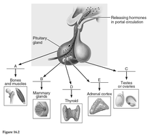
Key Terms:
Growth Hormone (GH): Stimulates growth of bones and muscles.
Prolactin (PRL): Stimulates milk production in mammary glands.
Follicle-Stimulating Hormone (FSH) / Luteinizing Hormone (LH): Stimulate gonads (testes/ovaries).
Thyroid-Stimulating Hormone (TSH): Stimulates the thyroid gland.
Adrenocorticotropic Hormone (ACTH): Stimulates the adrenal cortex.
Step-by-Step Guidance
Observe the diagram and the direction of each arrow from the pituitary gland to the target organ.
Recall which hormone targets each organ (e.g., GH to bones/muscles, TSH to thyroid, ACTH to adrenal cortex, etc.).
Match each label (A-E) to the correct hormone based on its target.
Write down your answers for each label before checking the final answer.
Try solving on your own before revealing the answer!
Final Answer:
A: Growth Hormone (GH)
B: Prolactin (PRL)
C: Follicle-Stimulating Hormone (FSH) / Luteinizing Hormone (LH)
D: Thyroid-Stimulating Hormone (TSH)
E: Adrenocorticotropic Hormone (ACTH)
Each hormone is released by the anterior pituitary and acts on a specific target organ to regulate important physiological processes.
