 Back
BackBIO 305 Study Guide: Neurons and Nervous System Organization
Study Guide - Smart Notes
 Tailored notes based on your materials, expanded with key definitions, examples, and context.
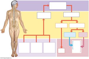
Q1. Label the structural and functional divisions of the nervous system using the provided diagram.
Background
Topic: Organization of the Nervous System
This question tests your understanding of how the nervous system is divided into central and peripheral components, and how these divisions relate to sensory and motor functions.
Key Terms and Concepts:
Central Nervous System (CNS): Consists of the brain and spinal cord; responsible for integrating and processing information.
Peripheral Nervous System (PNS): Includes all neural tissue outside the CNS; transmits sensory and motor signals.
Functional Divisions: Sensory (afferent) division, Motor (efferent) division, Somatic nervous system, Autonomic nervous system (with sympathetic and parasympathetic subdivisions).
Step-by-Step Guidance
Identify the two main anatomical divisions: the CNS (brain and spinal cord) and the PNS (all other neural elements).
Within the PNS, distinguish between the sensory (afferent) division, which brings information to the CNS, and the motor (efferent) division, which carries commands from the CNS to effectors.
Further divide the motor (efferent) division into the somatic nervous system (controls voluntary muscles) and the autonomic nervous system (controls involuntary functions).
Within the autonomic nervous system, identify the sympathetic and parasympathetic subdivisions, each with distinct roles in regulating body functions.
Try solving on your own before revealing the answer!
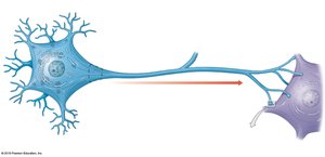
Q2. Label the parts of a neuron: dendrites, soma, nucleus, axon hillock, axon, axon terminal, arborizations, terminal boutons, pre-synaptic neuron, post-synaptic neuron.
Background
Topic: Neuron Structure and Function
This question assesses your ability to identify the main structural components of a neuron and understand their roles in neural signaling.
Key Terms:
Dendrites: Receive incoming signals.
Soma (cell body): Contains the nucleus and integrates signals.
Axon hillock: Initiates action potentials.
Axon: Conducts electrical impulses away from the soma.
Axon terminal/terminal boutons: Release neurotransmitters to communicate with other cells.
Pre-synaptic neuron: Sends the signal.
Post-synaptic neuron: Receives the signal.
Step-by-Step Guidance
Locate the dendrites, which are typically branching structures extending from the soma.
Identify the soma (cell body), which contains the nucleus.
Find the axon hillock, the region where the soma transitions into the axon.
Trace the axon, a long projection that carries signals away from the soma toward the axon terminals.
At the end of the axon, identify the axon terminals (terminal boutons), which form synapses with the next cell (post-synaptic neuron).
Try solving on your own before revealing the answer!
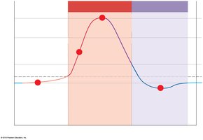
Q3. Interpret the graph of an action potential. What is happening during each time period? List the specific activities and channels involved.
Background
Topic: Action Potentials in Neurons
This question tests your ability to analyze the phases of an action potential and relate them to ion channel activity and membrane voltage changes.
Key Terms and Concepts:
Depolarization: Membrane potential becomes more positive, usually due to Na+ influx.
Repolarization: Membrane potential returns toward resting value, often due to K+ efflux.
Hyperpolarization: Membrane potential becomes more negative than resting potential.
Voltage-gated Na+ and K+ channels: Open and close at specific voltages to generate the action potential.
Step-by-Step Guidance
Identify the resting membrane potential (baseline before the spike).
Observe the rapid rise (depolarization) and note when voltage-gated Na+ channels open, allowing Na+ to enter the cell.
At the peak, voltage-gated Na+ channels inactivate and voltage-gated K+ channels open, starting repolarization.
During repolarization, K+ exits the cell, bringing the membrane potential back toward resting.
Hyperpolarization occurs as K+ channels remain open briefly, making the membrane potential more negative than the resting value.
Try solving on your own before revealing the answer!
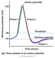
Q4. Draw and label an action potential graph for the average neuron. Label depolarization, repolarization, hyperpolarization, resting membrane potential, and threshold. Identify when sodium and potassium channels open and close.
Background
Topic: Action Potential Phases and Ion Channel Dynamics
This question requires you to understand and illustrate the sequence of events during an action potential, including the roles of Na+ and K+ channels.
Key Terms and Concepts:
Threshold: The critical level to which the membrane potential must be depolarized to initiate an action potential.
Resting membrane potential: The steady-state voltage across the membrane when the neuron is not firing.
Depolarization, repolarization, hyperpolarization: Key phases of the action potential.
Voltage-gated Na+ channels: Open rapidly at threshold, then inactivate.
Voltage-gated K+ channels: Open more slowly, contributing to repolarization and hyperpolarization.
Step-by-Step Guidance
Draw the baseline (resting membrane potential) and mark the threshold level.
Indicate the depolarization phase, where voltage-gated Na+ channels open and Na+ enters the cell.
Mark the peak of the action potential, where Na+ channels inactivate and K+ channels begin to open.
Show the repolarization phase, where K+ exits the cell, bringing the membrane potential back down.
Label the hyperpolarization phase, where K+ channels remain open briefly before returning to resting potential.
Try solving on your own before revealing the answer!
