Back
BackBIO163 Endocrine, Eye, and Ear Worksheet Study Guidance
Study Guide - Smart Notes
 Tailored notes based on your materials, expanded with key definitions, examples, and context.
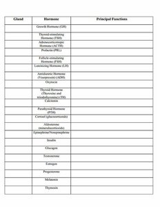
Q1. Complete the table: List the gland, hormone, principal function, and type (protein/steroid) for each hormone.
Background
Topic: Endocrine System
This question tests your knowledge of the major endocrine glands, the hormones they secrete, the main functions of these hormones, and whether each hormone is a protein or steroid type. Understanding these relationships is fundamental for studying hormone regulation and physiological effects in the body.
Key Terms and Concepts:
Endocrine gland: An organ that secretes hormones directly into the bloodstream.
Hormone: A chemical messenger that regulates physiological processes.
Principal function: The main effect or role of the hormone in the body.
Protein hormone: Hormones made of amino acids (e.g., insulin, growth hormone).
Steroid hormone: Hormones derived from cholesterol (e.g., cortisol, estrogen).
Step-by-Step Guidance
Start by identifying the gland associated with each hormone listed in the table (e.g., Growth Hormone is from the anterior pituitary).
For each hormone, recall or research its principal function (e.g., Growth Hormone stimulates growth and cell reproduction).
Determine whether each hormone is a protein or steroid based on its chemical structure or source (e.g., hormones from the adrenal cortex are typically steroids).
Fill in the table row by row, ensuring you match the hormone to the correct gland, function, and type.
Double-check your answers by reviewing textbook tables or lecture notes on endocrine hormones.
Try solving on your own before revealing the answer!
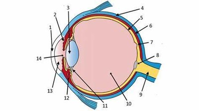
Q2. Eye Anatomy: Label the parts of the eye in the diagram and complete the table with structure, physiology/function, and location.
Background
Topic: Special Senses – Eye Anatomy and Physiology
This question assesses your ability to identify anatomical structures of the eye, describe their physiological roles, and specify their locations. This is essential for understanding how vision works and how different parts of the eye contribute to the process.
Key Terms and Concepts:
Cornea: Transparent front part of the eye that refracts light.
Retina: Layer containing photoreceptors (rods and cones) for vision.
Lens: Focuses light onto the retina.
Sclera: White, protective outer layer of the eye.
Optic nerve: Transmits visual information to the brain.
Step-by-Step Guidance
Examine the diagram and use your textbook or notes to identify each numbered structure (e.g., cornea, lens, retina, etc.).
For each structure, write a brief description of its main function (e.g., the cornea refracts light entering the eye).
Indicate the location of each structure (e.g., the retina lines the back of the eye).
Fill in the table with your findings, ensuring accuracy in both structure identification and function.
Review the diagram to confirm all parts are labeled correctly and correspond to your table entries.
Try solving on your own before revealing the answer!
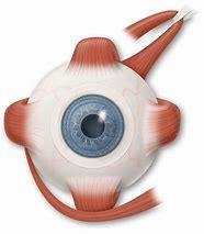
Q3. Label the external eye muscles, their functions, and innervation in the diagram.
Background
Topic: Eye Muscles and Innervation
This question tests your understanding of the six extraocular muscles, their actions (e.g., moving the eye up, down, laterally), and the cranial nerves that innervate them (III, IV, VI).
Key Terms and Concepts:
Extraocular muscles: Muscles that control eye movement (e.g., superior rectus, lateral rectus).
Innervation: The cranial nerve supplying each muscle (e.g., oculomotor nerve for most muscles).
Step-by-Step Guidance
Identify each muscle in the diagram (e.g., superior rectus, lateral rectus, etc.).
List the primary action of each muscle (e.g., superior rectus elevates the eye).
Determine which cranial nerve innervates each muscle (e.g., lateral rectus is innervated by cranial nerve VI).
Label the diagram and create a table or list summarizing muscle, function, and innervation.
Try solving on your own before revealing the answer!
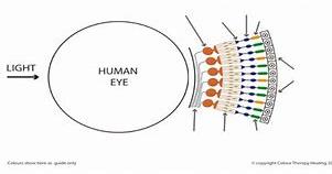
Q4. Label the photoreceptors in the diagram and list the function of each.
Background
Topic: Photoreceptors in the Retina
This question focuses on identifying rods and cones in the retina and understanding their roles in vision (rods for low light, cones for color and detail).
Key Terms and Concepts:
Rod cells: Photoreceptors sensitive to dim light, responsible for night vision.
Cone cells: Photoreceptors responsible for color vision and visual acuity.
Step-by-Step Guidance
Locate the layers of the retina in the diagram and identify where rods and cones are found.
Label each type of photoreceptor and briefly describe its function.
Note the distribution of rods and cones (e.g., cones are concentrated in the fovea).
Try solving on your own before revealing the answer!
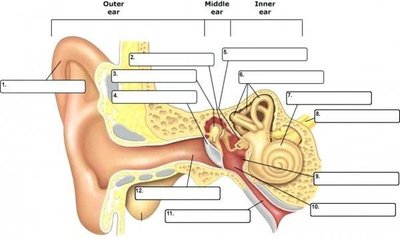
Q5. Label all structures in the ear diagram and list the function of each. Know the ossicles in order, organs for equilibrium and hearing, and the VIIIth cranial nerve divisions and function.
Background
Topic: Ear Anatomy and Physiology
This question tests your ability to identify the anatomical parts of the ear, understand the function of each part, and know the order of the ossicles, the organs responsible for static and dynamic equilibrium, hearing, and the divisions of the vestibulocochlear nerve (cranial nerve VIII).
Key Terms and Concepts:
Ossicles: Malleus, incus, stapes (in order from tympanic membrane to inner ear).
Static equilibrium: Sensed by the utricle and saccule.
Dynamic equilibrium: Sensed by the semicircular canals.
Hearing: Cochlea is the organ of hearing.
VIIIth cranial nerve: Vestibulocochlear nerve, with vestibular (balance) and cochlear (hearing) divisions.
Step-by-Step Guidance
Label each structure in the ear diagram (outer, middle, and inner ear components).
List the function of each labeled structure (e.g., tympanic membrane transmits sound vibrations).
Identify and order the ossicles (malleus, incus, stapes).
Indicate which structures are responsible for static and dynamic equilibrium, and for hearing.
Label the divisions of the vestibulocochlear nerve and describe their functions.
Try solving on your own before revealing the answer!