Back
BIO163 Exam 1 Study Guide: Human Body, Cells, Skin, Skeleton, and Muscles
Study Guide - Smart Notes
 Tailored notes based on your materials, expanded with key definitions, examples, and context.
Chapter 1: The Human Body – An Orientation
Medical Terminology
Understanding medical terms is essential for effective communication in anatomy and physiology. Medical terms are often derived from Latin or Greek roots and are used to describe body parts, functions, conditions, and procedures.
Prefix: Added to the beginning of a word to modify its meaning (e.g., hypo- means below).
Root: The main part of the word that contains its primary meaning (e.g., cardi means heart).
Suffix: Added to the end of a word to modify its meaning (e.g., -itis means inflammation).
Example: Gastritis = inflammation of the stomach.
Systems of the Body
The human body is organized into several systems, each with specific functions that contribute to overall homeostasis.
Integumentary System: Protects the body, regulates temperature, and provides sensory information.
Skeletal System: Provides support, protects organs, stores minerals, and produces blood cells.
Muscular System: Allows movement, maintains posture, and produces heat.
Nervous System: Controls body activities with electrical signals.
Endocrine System: Regulates body functions through hormones.
Cardiovascular System: Transports nutrients, gases, and wastes.
Lymphatic System: Defends against infection and disease.
Respiratory System: Facilitates gas exchange.
Digestive System: Breaks down food and absorbs nutrients.
Urinary System: Eliminates waste and maintains fluid balance.
Reproductive System: Produces offspring.
Body Planes, Quadrants, Regions, and Cavities
Body orientation is described using planes, quadrants, regions, and cavities to locate structures and describe relationships.
Planes: Sagittal (left/right), Frontal (anterior/posterior), Transverse (superior/inferior).
Quadrants: Right Upper, Left Upper, Right Lower, Left Lower (used for abdominal organs).
Regions: Nine regions (e.g., epigastric, hypogastric) for more precise localization.
Cavities: Dorsal (cranial, vertebral), Ventral (thoracic, abdominopelvic).
Chapter 3: Cells and Tissues
Structure of the Cell and Plasma Membrane
Cells are the basic units of life. The plasma membrane surrounds the cell, controlling the movement of substances in and out.
Plasma Membrane: Composed of a phospholipid bilayer with embedded proteins; selectively permeable.
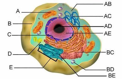
Organelles: Specialized structures within the cell (e.g., nucleus, mitochondria, endoplasmic reticulum).
DNA Properties and Protein Synthesis
DNA stores genetic information. Protein synthesis involves transcription and translation, using different types of RNA.
DNA: Double helix structure; contains genes that code for proteins.
Protein Synthesis:
Transcription: DNA is copied into messenger RNA (mRNA).
Translation: mRNA is decoded by ribosomes to assemble amino acids into proteins.
Types of RNA: mRNA (messenger), tRNA (transfer), rRNA (ribosomal).
Codons: Triplets of nucleotides on mRNA that specify amino acids.
Diffusion Laws and Plasma Membrane Selectivity
Diffusion is the movement of molecules from high to low concentration. The plasma membrane's selectivity regulates this process.
Concentration Gradient: Drives diffusion; molecules move down the gradient.
Selective Permeability: Only certain substances can cross the membrane freely.
Example: Oxygen and carbon dioxide diffuse across cell membranes.
Cell Shapes and Types of Tissues
Cells vary in shape and function. Tissues are groups of similar cells performing a common function.
Cell Shapes: Squamous (flat), cuboidal (cube-shaped), columnar (tall).
Types of Tissues: Epithelial, connective, muscle, nervous.
Cell Division (Mitosis)
Mitosis is the process by which cells divide to produce identical daughter cells, essential for growth and repair.
Phases: Prophase, metaphase, anaphase, telophase, cytokinesis.
Purpose: Growth, tissue repair, and asexual reproduction.
Genetic Code and Complementary Code
The genetic code is the set of rules by which information in DNA is translated into proteins. Complementary base pairing ensures accurate replication and transcription.
Genetic Code: Sequence of nucleotides in DNA and RNA that determines amino acid sequence.
Complementary Base Pairing: Adenine (A) pairs with Thymine (T), Cytosine (C) with Guanine (G) in DNA; in RNA, Uracil (U) replaces Thymine.
Chapter 4: Skin and Body Membranes
Skin Layers and Accessory Organs
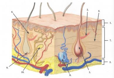
The skin is the largest organ, consisting of multiple layers and accessory structures such as glands and receptors.
Epidermis: Outermost layer; provides protection.
Dermis: Middle layer; contains blood vessels, nerves, glands, and hair follicles.
Hypodermis (Subcutaneous): Deepest layer; stores fat and anchors skin.
Accessory Organs: Sweat glands, sebaceous glands, hair follicles, nerve receptors.
Sweat Glands: Eccrine (all over body, regulate temperature), apocrine (armpits, groin, active after puberty).
Chapter 5: The Skeletal System
Bones of the Human Body – Skeleton and Articulations
The human skeleton provides structure, protection, and movement. Articulations (joints) connect bones and allow movement.
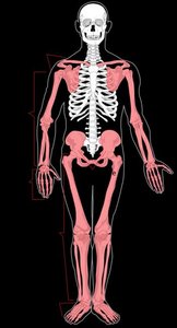
Axial Skeleton: Skull, vertebral column, rib cage.
Appendicular Skeleton: Limbs and girdles (shoulder and pelvic).
Articulations: Joints where two bones meet; classified by structure and function.
Associated Hormones and Bone Homeostasis
Bone homeostasis is regulated by hormones that control bone formation and resorption.
Parathyroid Hormone (PTH): Increases blood calcium by stimulating bone resorption.
Calcitonin: Lowers blood calcium by inhibiting bone resorption.
Growth Hormone: Stimulates bone growth during childhood.
Osteon Structure and Function
Osteons are the structural units of compact bone, providing strength and support.
Central Canal: Contains blood vessels and nerves.
Lamellae: Concentric rings of bone matrix.
Lacunae: Small spaces housing osteocytes.
Types of Joints and Joint Movements
Joints are classified by their structure and the movements they allow.
Fibrous Joints: Immovable (e.g., sutures of the skull).
Cartilaginous Joints: Slightly movable (e.g., intervertebral discs).
Synovial Joints: Freely movable (e.g., knee, shoulder).
Movements: Flexion, extension, abduction, adduction, rotation, circumduction.
Chapter 6: The Muscular System
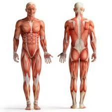
Muscles of the Human Body
Muscles are responsible for movement, posture, and heat production. The body contains over 600 muscles, classified as skeletal, cardiac, or smooth.
Skeletal Muscles: Voluntary, striated muscles attached to bones.
Cardiac Muscle: Involuntary, striated muscle found in the heart.
Smooth Muscle: Involuntary, non-striated muscle found in walls of organs.
Sarcomere Structure and Physiology of Contraction
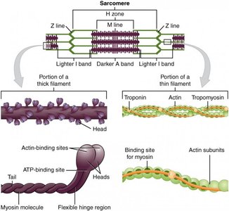
The sarcomere is the functional unit of muscle contraction, composed of actin and myosin filaments. Muscle contraction is triggered by ions and neurotransmitters.
Sarcomere: Bordered by Z lines; contains thick (myosin) and thin (actin) filaments.
Sliding Filament Theory: Myosin heads bind to actin, pulling filaments past each other to shorten the muscle.
Ions: Calcium ions (Ca2+) initiate contraction by binding to troponin.
Neurotransmitter: Acetylcholine (ACh) triggers muscle action potential.
Muscle Contraction Cycle
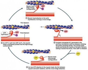
Muscle contraction involves a cyclical interaction between actin and myosin, powered by ATP hydrolysis.
Step 1: Myosin head attaches to actin, forming a cross-bridge.
Step 2: Power stroke: Myosin head pivots, pulling actin filament.
Step 3: ATP binds to myosin, causing detachment from actin.
Step 4: ATP hydrolysis re-cocks the myosin head for another cycle.