 Back
BackChapter 13 Guided Notes
Study Guide - Smart Notes
 Tailored notes based on your materials, expanded with key definitions, examples, and context.
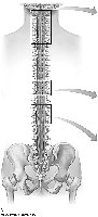
Q1. Label the spinal cord in the longitudinal view.
Background
Topic: Anatomy of the Spinal Cord (Longitudinal View)
This question tests your ability to identify and label the major anatomical regions and features of the spinal cord as seen from the side (longitudinal) view. Common structures include the cervical, thoracic, lumbar, and sacral regions, as well as the conus medullaris, cauda equina, and spinal nerves.
Key Terms:
Spinal cord regions: cervical, thoracic, lumbar, sacral
Conus medullaris
Cauda equina
Spinal nerves
Step-by-Step Guidance
Examine the longitudinal diagram and locate the main regions of the spinal cord (cervical, thoracic, lumbar, sacral).
Identify the conus medullaris, which is the tapered end of the spinal cord.
Find the cauda equina, which is the bundle of nerve roots extending below the conus medullaris.
Label the spinal nerves that branch off from each region.
Review the list of terms provided and match each to the correct anatomical location on the diagram.
Try solving on your own before revealing the answer!
Final Answer:
Cervical region: uppermost section, near the neck
Thoracic region: middle section, aligned with the ribcage
Lumbar region: lower back section
Sacral region: lowest section, near the pelvis
Conus medullaris: tapered end of the spinal cord, around L1-L2
Cauda equina: bundle of nerves extending below the conus medullaris
Spinal nerves: branch off laterally from each region
Label each structure on the diagram according to its anatomical position.
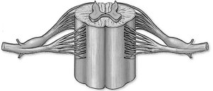
Q2. Label the cross-sectional view of the spinal cord with the list terms.
Background
Topic: Cross-Sectional Anatomy of the Spinal Cord
This question focuses on identifying the internal structures of the spinal cord as seen in a cross-section. Key features include gray matter, white matter, dorsal and ventral roots, and the central canal.
Key Terms:
Gray matter (dorsal horn, ventral horn, lateral horn)
White matter (funiculi/columns)
Central canal
Dorsal root and ventral root
Spinal nerve
Step-by-Step Guidance
Locate the butterfly-shaped gray matter in the center of the cross-section.
Identify the dorsal (posterior) and ventral (anterior) horns of the gray matter.
Find the surrounding white matter, which is organized into columns (funiculi).
Locate the central canal in the middle of the gray matter.
Identify the dorsal and ventral roots extending from the spinal cord, and where they merge to form the spinal nerve.
Try solving on your own before revealing the answer!
Final Answer:
Gray matter: central butterfly-shaped area (dorsal, ventral, lateral horns)
White matter: outer region surrounding gray matter (anterior, lateral, posterior funiculi)
Central canal: small hole in the center of gray matter
Dorsal root: posterior root carrying sensory fibers
Ventral root: anterior root carrying motor fibers
Spinal nerve: formed by the union of dorsal and ventral roots
Label each structure on the diagram according to its anatomical location.
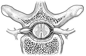
Q3. Label the meninges and spaces surrounding the spinal cord. What flows within the subarachnoid space?
Background
Topic: Meninges and Spaces of the Spinal Cord
This question tests your understanding of the protective coverings (meninges) of the spinal cord and the spaces between them. It also asks about the fluid found in the subarachnoid space.
Key Terms:
Dura mater
Arachnoid mater
Pia mater
Epidural space
Subdural space
Subarachnoid space
Cerebrospinal fluid (CSF)
Step-by-Step Guidance
Identify the three layers of meninges from outermost to innermost: dura mater, arachnoid mater, pia mater.
Locate the spaces between these layers: epidural space (outside dura), subdural space (between dura and arachnoid), subarachnoid space (between arachnoid and pia).
Label each meningeal layer and space on the diagram.
Recall what substance circulates within the subarachnoid space.
Try solving on your own before revealing the answer!
Final Answer:
Dura mater: outermost meningeal layer
Arachnoid mater: middle meningeal layer
Pia mater: innermost meningeal layer, directly on the spinal cord
Epidural space: between bone and dura mater
Subdural space: between dura mater and arachnoid mater
Subarachnoid space: between arachnoid mater and pia mater
Cerebrospinal fluid (CSF) flows within the subarachnoid space
CSF cushions and protects the spinal cord within the subarachnoid space.
Q4. Fill in the blank – PNS Organization
Background
Topic: Peripheral Nervous System (PNS) Structure and Function
This question tests your knowledge of the basic organization of the PNS, including its main components and the direction of information flow in sensory and motor pathways.
Key Terms:
PNS: Peripheral Nervous System
Nerves
Ganglia
Sensory (afferent) pathways
Motor (efferent) pathways
Step-by-Step Guidance
Recall the two main structural components of the PNS found outside the CNS.
Think about the direction in which sensory pathways carry information (toward or away from the CNS).
Consider the direction in which motor pathways carry information.
Fill in the blanks with the correct terms based on your understanding.
Try solving on your own before revealing the answer!
Final Answer:
The PNS consists of nerves and ganglia outside the CNS.
Sensory pathways carry information toward the CNS, while motor pathways carry information away from the CNS.
Nerves are bundles of axons, and ganglia are clusters of neuron cell bodies in the PNS.
Q5. Fill in the Blank – Spinal Nerves & Plexuses
Background
Topic: Spinal Nerves and Nerve Plexuses
This question tests your knowledge of the number of spinal nerve pairs and the names of the major nerve plexuses in the body.
Key Terms:
Spinal nerves
Plexus (plural: plexuses)
Cervical, brachial, lumbar, sacral plexuses
Step-by-Step Guidance
Recall the total number of pairs of spinal nerves in the human body.
List the four major nerve plexuses, starting with cervical.
Fill in the blanks with the correct numbers and names.
Try solving on your own before revealing the answer!
Final Answer:
There are 31 pairs of spinal nerves.
The major nerve plexuses include cervical, brachial, lumbar, and sacral.
Each plexus supplies nerves to specific regions of the body.
