 Back
BIO168 Unit 2 Study Guide: Integumentary and Skeletal Systems
Study Guide - Smart Notes
 Tailored notes based on your materials, expanded with key definitions, examples, and context.
Q1. List the skin layers in order.
Background
Topic: Integumentary System
This question tests your knowledge of the structural organization of the skin, which is essential for understanding its functions and clinical relevance.
Key Terms:
Epidermis
Dermis
Hypodermis (subcutaneous layer)
Step-by-Step Guidance
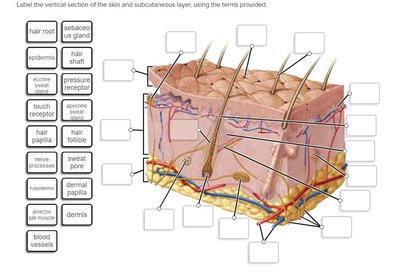
Recall that the skin is composed of three main layers, each with distinct functions and structures.
Start from the outermost layer and move inward, identifying each layer by its name and general characteristics.
Consider the order: which layer is exposed to the environment, which provides structural support, and which connects to underlying tissues?
Review the diagram for visual confirmation of the layers and their arrangement.
Try solving on your own before revealing the answer!
Final Answer:
Epidermis (outermost), Dermis (middle), Hypodermis (deepest/subcutaneous layer).
These layers are arranged from superficial to deep, each serving unique functions in protection, sensation, and insulation.
Q2. Describe all associated structures (sweat and sebum glands) and their functions.
Background
Topic: Skin Appendages
This question focuses on the accessory structures of the skin, specifically sweat glands and sebaceous (sebum) glands, and their physiological roles.
Key Terms:
Sweat glands (eccrine and apocrine)
Sebaceous glands
Functions: thermoregulation, excretion, lubrication
Step-by-Step Guidance
Identify the two main types of sweat glands: eccrine and apocrine.
Describe the location and function of each gland type (eccrine: widespread, apocrine: specific areas).
Explain the role of sebaceous glands in producing sebum and their association with hair follicles.
Relate each gland's function to overall skin health and homeostasis.
Try solving on your own before revealing the answer!
Final Answer:
Eccrine glands produce watery sweat for cooling; apocrine glands produce thicker sweat in specific areas; sebaceous glands secrete sebum to lubricate skin and hair.
These structures help regulate temperature, excrete waste, and maintain skin integrity.
Q3. List the epidermis layers in order.
Background
Topic: Epidermal Structure
This question tests your knowledge of the stratified arrangement of the epidermis, which is crucial for understanding skin function and regeneration.
Key Terms:
Stratum corneum
Stratum lucidum (only in thick skin)
Stratum granulosum
Stratum spinosum
Stratum basale
Step-by-Step Guidance
Recall the five layers of the epidermis, starting from the surface and moving inward.
Note that the stratum lucidum is only present in thick skin (palms, soles).
Arrange the layers in order, considering their function and location.
Use the diagram to visualize the layers and their relative positions.
Try solving on your own before revealing the answer!
Final Answer:
Stratum corneum, stratum lucidum (if present), stratum granulosum, stratum spinosum, stratum basale.
These layers are arranged from superficial to deep, each contributing to skin protection and regeneration.
Q4. Explain the degrees of burns and the rule of nines.
Background
Topic: Skin Pathology
This question assesses your understanding of burn classification and the clinical method for estimating burn area.
Key Terms:
First, second, third degree burns
Rule of nines
Step-by-Step Guidance
Define each degree of burn based on depth and tissue involvement.
Describe the clinical significance of each burn type.
Explain the rule of nines for estimating total body surface area affected by burns.
Apply the rule to hypothetical scenarios for practice.
Try solving on your own before revealing the answer!
Final Answer:
First degree: epidermis only; second degree: epidermis and part of dermis; third degree: full thickness. Rule of nines divides the body into regions, each representing 9% (or multiples) of total surface area.
This helps clinicians quickly assess severity and plan treatment.
Q5. Discuss skin color, pigments, and related pathologies.
Background
Topic: Skin Pigmentation
This question explores the biological basis of skin color and common disorders associated with pigmentation.
Key Terms:
Melanin, carotene, hemoglobin
Pathologies: albinism, jaundice, cyanosis
Step-by-Step Guidance
Identify the main pigments responsible for skin color.
Describe how each pigment contributes to overall skin tone.
Discuss common pathologies and their clinical presentation.
Relate pigment changes to underlying health conditions.
Try solving on your own before revealing the answer!
Final Answer:
Melanin (brown/black), carotene (yellow/orange), hemoglobin (red). Pathologies include albinism (lack of melanin), jaundice (yellow from bilirubin), cyanosis (blue from low oxygen).
These changes can indicate underlying health issues.
Q6. Describe the structure of spongy bone.
Background
Topic: Skeletal System
This question tests your understanding of the microscopic and macroscopic structure of spongy bone and its functional significance.
Key Terms:
Trabeculae
Bone marrow
Osteocytes
Step-by-Step Guidance
Define spongy bone and its location within bones.
Describe the trabecular network and its role in strength and weight reduction.
Explain the presence of bone marrow and its function.
Relate the structure to bone physiology and hematopoiesis.
Try solving on your own before revealing the answer!
Final Answer:
Spongy bone consists of a lattice of trabeculae, spaces filled with bone marrow, and osteocytes within lacunae.
This structure provides strength and supports blood cell formation.
Q7. Identify all structures in the long bone diagram.
Background
Topic: Long Bone Anatomy
This question requires you to recognize and label the anatomical features of a long bone, which is fundamental for understanding bone function and pathology.
Key Terms:
Diaphysis, epiphysis, metaphysis
Periosteum, endosteum
Medullary cavity, articular cartilage
Step-by-Step Guidance
Review the diagram and identify the main regions of the bone.
Label each structure based on its location and function.
Consider the relationship between these structures and bone growth or repair.
Use the diagram to reinforce your understanding of spatial arrangement.
Try solving on your own before revealing the answer!
Final Answer:
Diaphysis (shaft), epiphysis (ends), metaphysis (growth region), periosteum (outer covering), endosteum (inner lining), medullary cavity (center), articular cartilage (joint surface).
Each structure plays a role in bone growth, protection, and movement.
Q8. Explain bone growth and development: Types of ossification.
Background
Topic: Bone Development
This question tests your understanding of the processes by which bones form and grow, including intramembranous and endochondral ossification.
Key Terms:
Intramembranous ossification
Endochondral ossification
Step-by-Step Guidance
Define ossification and its importance in bone development.
Describe intramembranous ossification and its role in forming flat bones.
Explain endochondral ossification and its role in forming long bones.
Compare and contrast the two processes.
Try solving on your own before revealing the answer!
Final Answer:
Intramembranous ossification forms flat bones from mesenchyme; endochondral ossification forms long bones from cartilage templates.
Both processes are essential for skeletal development and growth.
Q9. Describe bone homeostasis: organic/inorganic matrix.
Background
Topic: Bone Physiology
This question explores the balance between organic and inorganic components in bone, which is crucial for strength and flexibility.
Key Terms:
Organic matrix: collagen fibers
Inorganic matrix: hydroxyapatite (calcium phosphate)
Step-by-Step Guidance
Define bone homeostasis and its importance.
Describe the composition and function of the organic matrix.
Explain the role of the inorganic matrix in bone strength.
Discuss how these components interact to maintain bone health.
Try solving on your own before revealing the answer!
Final Answer:
Organic matrix provides flexibility; inorganic matrix gives strength. Homeostasis maintains the balance for optimal bone function.
Disruption can lead to bone diseases.
Q10. List hormones associated with bone homeostasis.
Background
Topic: Endocrine Regulation of Bone
This question tests your knowledge of hormonal control over bone remodeling and calcium balance.
Key Terms:
Parathyroid hormone (PTH)
Calcitonin
Vitamin D
Step-by-Step Guidance
Identify the main hormones involved in bone homeostasis.
Describe the effect of each hormone on bone and calcium levels.
Explain the feedback mechanisms regulating these hormones.
Relate hormonal imbalances to bone disorders.
Try solving on your own before revealing the answer!
Final Answer:
PTH increases blood calcium by stimulating bone resorption; calcitonin lowers blood calcium by inhibiting resorption; vitamin D enhances calcium absorption.
These hormones work together to maintain bone and mineral balance.
Q11. Name the shapes of bones.
Background
Topic: Bone Classification
This question tests your ability to classify bones based on their shapes, which is important for anatomical identification.
Key Terms:
Long, short, flat, irregular, sesamoid bones
Step-by-Step Guidance
List the five main bone shapes.
Provide examples of each type.
Explain the functional significance of each shape.
Relate bone shape to location and movement.
Try solving on your own before revealing the answer!
Final Answer:
Long (femur), short (carpals), flat (skull), irregular (vertebrae), sesamoid (patella).
Each shape is adapted for specific functions.
Q12. Identify cranial bones and their markings.
Background
Topic: Skull Anatomy
This question tests your knowledge of the bones forming the cranium and their anatomical features.
Key Terms:
Frontal, parietal, temporal, occipital, sphenoid, ethmoid bones
Bone markings: foramina, processes, sutures
Step-by-Step Guidance
List the cranial bones.
Identify key markings on each bone.
Explain the function of these markings (e.g., passage of nerves, muscle attachment).
Relate markings to clinical or anatomical significance.
Try solving on your own before revealing the answer!
Final Answer:
Frontal, parietal, temporal, occipital, sphenoid, ethmoid; markings include foramina (holes), processes (projections), and sutures (joints).
These features are important for protection and function.
Q13. Define sutures.
Background
Topic: Skull Joints
This question tests your understanding of the fibrous joints connecting cranial bones.
Key Terms:
Sutures
Fibrous joint
Step-by-Step Guidance
Define what a suture is in anatomical terms.
Describe the role of sutures in skull development and protection.
Identify major sutures (e.g., coronal, sagittal, lambdoid).
Relate sutures to clinical conditions (e.g., craniosynostosis).
Try solving on your own before revealing the answer!
Final Answer:
Sutures are immovable fibrous joints between skull bones, allowing growth and protection.
Major sutures include coronal, sagittal, lambdoid, and squamous.
Q14. Name all axial and appendicular skeletal bones.
Background
Topic: Skeletal System Organization
This question tests your ability to distinguish between the bones of the axial and appendicular skeleton.
Key Terms:
Axial skeleton: skull, vertebral column, rib cage
Appendicular skeleton: limbs, girdles
Step-by-Step Guidance
List the bones included in the axial skeleton.
List the bones included in the appendicular skeleton.
Explain the functional differences between the two divisions.
Relate bone grouping to movement and protection.
Try solving on your own before revealing the answer!
Final Answer:
Axial: skull, vertebral column, rib cage; appendicular: limbs, shoulder girdle, pelvic girdle.
Axial provides support and protection; appendicular enables movement.
Q15. Compare shoulder and pelvic girdles: male vs. female.
Background
Topic: Skeletal Anatomy
This question tests your understanding of the structural differences between male and female girdles and their functional implications.
Key Terms:
Shoulder girdle: clavicle, scapula
Pelvic girdle: ilium, ischium, pubis
Sexual dimorphism
Step-by-Step Guidance
Identify the bones forming each girdle.
Describe the general differences between male and female pelvic girdles (e.g., width, shape).
Explain the functional significance of these differences (e.g., childbirth).
Compare shoulder girdle features if relevant.
Try solving on your own before revealing the answer!
Final Answer:
Female pelvic girdle is wider and more circular for childbirth; male is narrower and more robust. Shoulder girdle differences are less pronounced.
These adaptations reflect functional needs.
Q16. Differentiate types of vertebrae and spine curvatures.
Background
Topic: Vertebral Column Anatomy
This question tests your ability to distinguish between cervical, thoracic, lumbar, sacral, and coccygeal vertebrae and understand normal and abnormal curvatures.
Key Terms:
Cervical, thoracic, lumbar, sacral, coccygeal vertebrae
Curvatures: lordosis, kyphosis, scoliosis
Step-by-Step Guidance
List the five types of vertebrae and their distinguishing features.
Describe normal spinal curvatures and their functions.
Explain abnormal curvatures and their clinical significance.
Relate vertebral structure to movement and protection.
Try solving on your own before revealing the answer!
Final Answer:
Cervical (neck), thoracic (chest), lumbar (lower back), sacral (pelvis), coccygeal (tailbone); curvatures include lordosis (inward), kyphosis (outward), scoliosis (lateral).
Each type and curvature has specific functions and clinical implications.
Q17. Describe bone fractures and the healing process.
Background
Topic: Bone Injury and Repair
This question tests your understanding of fracture types and the physiological steps involved in bone healing.
Key Terms:
Fracture types: simple, compound, comminuted, greenstick
Healing stages: hematoma, callus formation, remodeling
Step-by-Step Guidance
Identify common types of bone fractures.
Describe the sequence of events in bone healing.
Explain the role of cells and tissues in each stage.
Relate healing to clinical management.
Try solving on your own before revealing the answer!
Final Answer:
Fractures include simple, compound, comminuted, greenstick; healing involves hematoma, callus formation, and remodeling.
Each stage is essential for restoring bone integrity.
Q18. Classify joints: functional and structural classification.
Background
Topic: Joint Anatomy
This question tests your ability to classify joints based on movement and structure.
Key Terms:
Functional: synarthrosis, amphiarthrosis, diarthrosis
Structural: fibrous, cartilaginous, synovial
Step-by-Step Guidance
Define functional classification based on movement.
Define structural classification based on tissue type.
Provide examples of each classification.
Relate classification to joint function and location.
Try solving on your own before revealing the answer!
Final Answer:
Functional: synarthrosis (immovable), amphiarthrosis (slightly movable), diarthrosis (freely movable); structural: fibrous, cartilaginous, synovial.
Classification helps predict movement and stability.
Q19. Describe types of joint movements.
Background
Topic: Joint Physiology
This question tests your knowledge of the various movements possible at different joints.
Key Terms:
Flexion, extension, abduction, adduction, rotation, circumduction
Step-by-Step Guidance
List the main types of joint movements.
Define each movement and provide examples.
Relate movement type to joint structure.
Use diagrams or models to visualize movements.
Try solving on your own before revealing the answer!
Final Answer:
Movements include flexion, extension, abduction, adduction, rotation, circumduction; each occurs at specific joints.
Understanding movement types aids in clinical assessment.
Q20. Describe the anatomical structure of a synovial joint.
Background
Topic: Synovial Joint Anatomy
This question tests your ability to identify and describe the components of a synovial joint, which is essential for understanding joint function and pathology.
Key Terms:
Articular cartilage
Joint cavity
Synovial membrane
Fibrous capsule
Ligaments
Step-by-Step Guidance
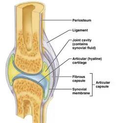
Identify the main components of a synovial joint.
Describe the function of each component (e.g., articular cartilage reduces friction).
Explain how these structures work together to allow movement and stability.
Use the diagram to visualize the arrangement and relationships of the components.
Try solving on your own before revealing the answer!
Final Answer:
Synovial joints have articular cartilage, joint cavity with synovial fluid, synovial membrane, fibrous capsule, and ligaments.
These structures enable free movement and protect joint surfaces.
