 Back
BackBIO200 Study Guide: Anatomy, Histology, and Integumentary System
Study Guide - Smart Notes
 Tailored notes based on your materials, expanded with key definitions, examples, and context.
Anatomic Position and Planes
Anatomic Position
The anatomic position is a standardized posture used as a reference in anatomy. The body stands upright, facing forward, arms at the sides with palms facing forward, and feet together. This position ensures consistency when describing locations and directions on the body.
Anatomic Planes
Anatomic planes are imaginary lines used to divide the body:
Coronal (Frontal) Plane: Divides the body into anterior (front) and posterior (back) sections.
Sagittal Plane: Divides the body into left and right sections. The midsagittal plane divides it exactly at the midline.
Transverse (Horizontal) Plane: Divides the body into superior (upper) and inferior (lower) sections.
Directional Terms
Directional terms describe the location of structures relative to others:
Anterior: Toward the front
Posterior: Toward the back
Superior: Above
Inferior: Below
Lateral: Away from the midline
Medial: Toward the midline
Distal: Farther from the point of attachment
Proximal: Closer to the point of attachment
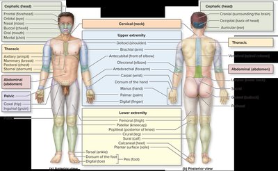
Regional Terms
Regional terms specify areas of the body, such as cephalic (head), cervical (neck), thoracic (chest), abdominal (abdomen), pelvic (pelvis), and extremities.
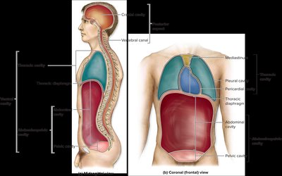
Body Cavities
Major Body Cavities
The body contains several cavities that house organs:
Dorsal cavity: Includes the cranial cavity (brain) and vertebral canal (spinal cord).
Ventral cavity: Includes the thoracic cavity (heart, lungs), abdominopelvic cavity (digestive organs, reproductive organs).
Thoracic cavity: Contains the pleural cavities (lungs), pericardial cavity (heart), and mediastinum.
Abdominal cavity: Contains digestive organs.
Pelvic cavity: Contains urinary bladder, reproductive organs.
Abdominopelvic Regions & Quadrants
The abdominopelvic area is divided into regions and quadrants for clinical reference. Each quadrant contains specific organs:
Right Upper Quadrant (RUQ): Liver, gallbladder
Left Upper Quadrant (LUQ): Stomach, spleen
Right Lower Quadrant (RLQ): Appendix, cecum
Left Lower Quadrant (LLQ): Descending colon, sigmoid colon
Histology: Tissues
Types of Tissues
There are four primary tissue types in the human body:
Epithelial Tissue: Covers surfaces, lines cavities, forms glands.
Connective Tissue: Supports, protects, and binds other tissues.
Muscle Tissue: Contracts to produce movement.
Nervous Tissue: Conducts electrical impulses for communication.
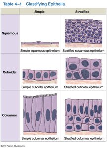
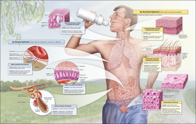
Epithelial Tissue Classification
Epithelial tissues are classified by cell layers and cell shape:
Cell Layers:
Simple: Single layer
Stratified: Multiple layers
Cell Shape:
Squamous: Flat
Cuboidal: Cube-shaped
Columnar: Column-shaped
Simple | Stratified | |
|---|---|---|
Squamous | Simple squamous epithelium | Stratified squamous epithelium |
Cuboidal | Simple cuboidal epithelium | Stratified cuboidal epithelium |
Columnar | Simple columnar epithelium | Stratified columnar epithelium |
Connective Tissue Classification
Connective tissues are classified based on their matrix and cell arrangement:
Connective Tissue Proper:
Loose (areolar, adipose)
Dense (fibrous)
Fluid Connective Tissue:
Blood
Lymph
Supporting Connective Tissue:
Cartilage
Bone
Integumentary System
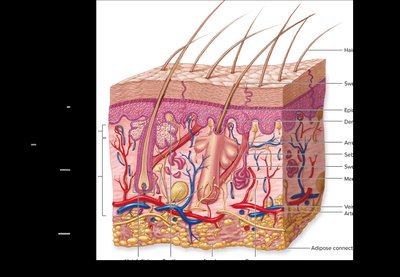
Layers of the Skin
The skin consists of two main layers:
Epidermis: The outermost layer, composed of stratified squamous epithelium. It provides protection and contains several sublayers (stratum basale, stratum spinosum, stratum granulosum, stratum lucidum, stratum corneum).
Dermis: The deeper layer, composed of connective tissue. It contains blood vessels, nerves, hair follicles, sweat glands, and other structures.
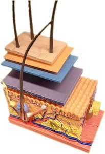
Skin Lab Model
Skin models are used in practical labs to identify the layers and structures of the skin, including hair follicles, glands, and blood vessels.
Other Structures within the Dermis
Hair follicles
Sweat glands
Sebaceous glands
Blood vessels
Nerve endings
Microscopic Identification of Tissues
Identifying Epithelial and Connective Tissues
Students should be able to identify epithelial and connective tissues under the microscope based on cell shape, arrangement, and matrix characteristics.
Examples
Simple squamous epithelium: Found in alveoli of lungs
Stratified squamous epithelium: Found in skin
Loose connective tissue: Found under epithelia
Dense connective tissue: Found in tendons
Summary Table: Tissue Types and Functions
Tissue Type | Main Function | Example Location |
|---|---|---|
Epithelial | Protection, absorption, secretion | Skin, lining of GI tract |
Connective | Support, binding, transport | Tendons, blood |
Muscle | Movement | Skeletal muscles, heart |
Nervous | Communication | Brain, nerves |
Additional info: Expanded explanations and examples were added for completeness and academic context.
