 Back
BackBIOL 2113 Final Exam Review – Step-by-Step Study Guidance
Study Guide - Smart Notes
 Tailored notes based on your materials, expanded with key definitions, examples, and context.
Q1. List and describe the layers of the epidermis from superficial to deep.
Background
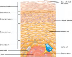
Topic: Integumentary System – Skin Structure
This question tests your understanding of the organization and function of the epidermal layers, which is essential for understanding skin physiology and pathology.
Key Terms:
Stratum corneum: Outermost layer, composed of dead, keratin-filled cells.
Stratum lucidum: Thin, clear layer found only in thick skin (palms, soles).
Stratum granulosum: Contains granules; cells begin to die and keratinize.
Stratum spinosum: Several layers of keratinocytes; provides strength and flexibility.
Stratum basale: Deepest layer; site of cell division and melanocyte location.
Step-by-Step Guidance
Start by identifying the most superficial layer of the epidermis and describe its main characteristics (e.g., cell type, function).
Move to the next deeper layer and note any unique features or functions (such as presence of granules or clear cells).
Continue this process for each layer, making sure to describe the transition from dead, keratinized cells at the surface to living, dividing cells at the base.
Consider the role of specialized cells (like melanocytes and Merkel cells) and where they are located within these layers.
Try solving on your own before revealing the answer!
Final Answer:
From superficial to deep, the layers are:
Stratum corneum: Dead, keratin-filled cells that provide a tough, protective barrier.
Stratum lucidum: Thin, clear layer found only in thick skin; provides extra protection.
Stratum granulosum: Cells contain keratohyalin granules; begin to die and keratinize.
Stratum spinosum: Several layers of keratinocytes connected by desmosomes; provides strength.
Stratum basale: Single row of dividing cells; contains melanocytes and Merkel cells.
This organization allows for continuous renewal and protection of the skin.
Q2. Describe the structure of long bones and identify the major regions and components.
Background
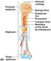
Topic: Skeletal System – Bone Structure
This question assesses your knowledge of the anatomy of long bones, which is foundational for understanding bone growth, repair, and function.
Key Terms:
Diaphysis: Shaft of the bone, mainly compact bone.
Epiphyses: Ends of the bone, mostly spongy bone.
Metaphysis: Region between diaphysis and epiphysis; includes growth plate in children.
Articular cartilage: Covers joint surfaces, reduces friction.
Medullary cavity: Hollow center containing bone marrow.
Periosteum: Outer covering, contains blood supply and growth cells.
Endosteum: Inner lining of the medullary cavity.
Step-by-Step Guidance
Begin by identifying the main shaft of the bone and describe its composition and function.
Locate the ends of the bone and explain how their structure differs from the shaft.
Describe the region between the shaft and the ends, noting its importance in bone growth.
Identify the coverings and linings of the bone, and explain their roles in nourishment, growth, and repair.
Try solving on your own before revealing the answer!
Final Answer:
The major regions and components of a long bone are:
Diaphysis: The shaft, composed mainly of compact bone for strength.
Epiphyses: The ends, containing spongy bone and red marrow.
Metaphysis: The region between diaphysis and epiphysis, including the growth plate in children.
Articular cartilage: Covers joint surfaces to reduce friction.
Medullary cavity: Central cavity containing yellow marrow (fat storage).
Periosteum: Tough outer membrane for protection and attachment of tendons/ligaments.
Endosteum: Thin membrane lining the medullary cavity, involved in bone remodeling.
This structure supports bone growth, repair, and function.
