 Back
BackBIOL 71 Lab Practical Study Guide: Cell, Tissue, and Integumentary System Essentials
Study Guide - Smart Notes
 Tailored notes based on your materials, expanded with key definitions, examples, and context.
Cell Biology
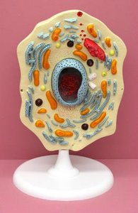
Cell Structure and Organelles
The cell is the fundamental unit of life, composed of various organelles that perform specialized functions. Understanding cell structure is essential for studying anatomy and physiology.
Plasma Membrane: The boundary of the cell, regulates entry and exit of substances.
Microvilli: Finger-like projections that increase surface area for absorption.
Cilia: Hair-like structures for movement of substances across cell surfaces.
Flagella: Tail-like structures for cell movement.
Cytoplasm: The region between the plasma membrane and nucleus, containing cytosol and organelles.
Nucleus: Contains genetic material (DNA); controls cell activities.
Nucleolus: Site of ribosome synthesis within the nucleus.
Nuclear Pores: Openings in the nuclear envelope for transport of molecules.
Chromatin: DNA and protein complex; condenses to form chromosomes during cell division.
Endoplasmic Reticulum (ER):
Rough ER: Studded with ribosomes; synthesizes proteins.
Smooth ER: Lacks ribosomes; synthesizes lipids and detoxifies chemicals.
Ribosome: Site of protein synthesis.
Golgi Apparatus: Modifies, sorts, and packages proteins and lipids for secretion or use within the cell.
Cytoskeleton: Network of protein filaments for cell shape and movement.
Secretory Vesicle: Transports substances out of the cell.
Peroxisome: Breaks down fatty acids and detoxifies harmful substances.
Lysosome: Contains digestive enzymes for breaking down waste.
Mitochondria: Powerhouse of the cell; site of ATP production.
Example: The mitochondria generate energy for muscle contraction.
Microscopy
Microscope Parts and Function
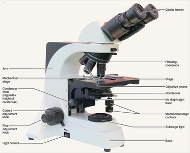
Microscopes are essential tools for studying cells and tissues. Understanding their parts and operation is crucial for laboratory work.
Ocular Lens: Eyepiece for viewing specimens.
Objective Lens: Provides different magnifications (4X, 10X, 40X, 100X).
Stage: Platform where slides are placed.
Coarse Adjustment: Moves stage for focusing at low magnification.
Fine Adjustment: Fine-tunes focus at higher magnification.
Total Magnification: Product of ocular and objective lens magnifications.
Field of View: Area visible through the microscope.
Resolution: Ability to distinguish two close objects as separate.
Example: Using the oil immersion objective (100X) increases resolution for viewing bacteria.
Tissue Types
Classification and Examples
Tissues are groups of cells with similar structure and function. The four main types are epithelial, connective, muscle, and nervous tissue.
Epithelial Tissue: Covers surfaces, lines cavities, forms glands.
Connective Tissue: Supports, protects, and binds other tissues. Includes blood, bone, cartilage, areolar, dense, and adipose tissues.
Muscle Tissue: Responsible for movement. Types: skeletal, cardiac, smooth.
Nervous Tissue: Conducts electrical impulses; found in brain, spinal cord, nerves.
Blood
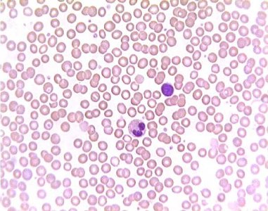
Blood is a fluid connective tissue composed of cells suspended in plasma. It transports oxygen, nutrients, and waste.
Bone
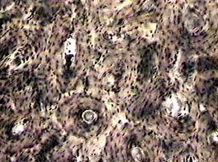
Bone tissue provides structural support and protection. It consists of osteocytes in lacunae, surrounded by a mineralized matrix.
Cartilage
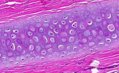
Cartilage is a flexible connective tissue with chondrocytes in lacunae, found in joints, ear, nose, and respiratory tract.
Adipose Tissue
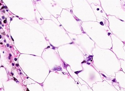
Adipose tissue stores fat and provides insulation and protection.
Muscle Tissue
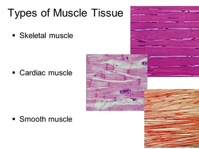
Muscle tissue is specialized for contraction. There are three types: skeletal (voluntary movement), cardiac (heart), and smooth (walls of organs).
Skeletal Muscle: Striated, voluntary.
Cardiac Muscle: Striated, involuntary, intercalated discs.
Smooth Muscle: Non-striated, involuntary.
Epithelial Tissues
Types and Associated Structures
Epithelial tissues are classified by cell shape and layers. They line surfaces and cavities, and form glands.
Simple Squamous: Single layer of flat cells; found in alveoli, blood vessels.
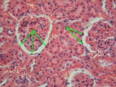
Simple Cuboidal: Single layer of cube-shaped cells; found in kidney tubules.
Simple Columnar: Single layer of tall cells; found in digestive tract.
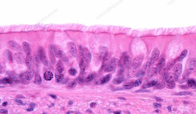
Ciliated Pseudostratified Columnar: Appears layered, but all cells touch basement membrane; found in respiratory tract.
Stratified Squamous: Multiple layers; protects against abrasion; found in skin, mouth.
Stratified Cuboidal/Columnar: Rare; found in ducts of glands.
Cilia: Move substances across surface.
Apical Surface: Exposed to lumen or external environment.
Basal Surface: Attached to basement membrane.
Basement Membrane: Supports epithelium.
Lumen: Internal space of a tubular structure.
Simple Squamous Epithelium
Simple Cuboidal Epithelium
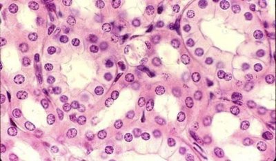
Simple Columnar Epithelium
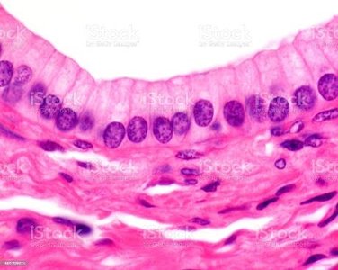
Stratified Squamous Epithelium
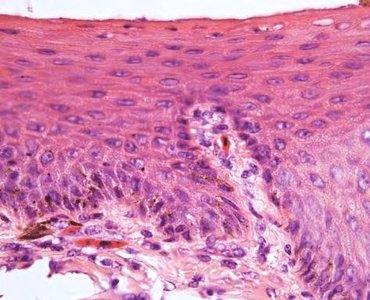
Stratified Columnar Epithelium
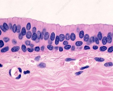
Pseudostratified Ciliated Columnar Epithelium
The Integumentary System
Skin Structure and Accessory Structures
The skin is the largest organ, providing protection, sensation, and regulation. It consists of multiple layers and accessory structures.
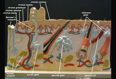
Epidermis: Outermost layer; composed of stratified squamous epithelium.
Dermis: Middle layer; contains connective tissue, blood vessels, nerves.
Papillary Layer: Superficial dermis; contains capillaries and nerve endings.
Reticular Layer: Deep dermis; dense connective tissue.
Hypodermis (SubQ layer): Deepest layer; adipose tissue.
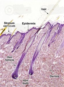
Hair Shaft/Follicle: Produces hair.
Arrector Pili Muscle: Causes hair to stand up.
Sebaceous Gland: Secretes oil (sebum).
Sweat Gland: Produces sweat for cooling.
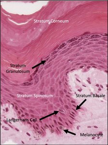
Stratum Basale: Deepest epidermal layer; mitotic activity.
Stratum Spinosum: Provides strength and flexibility.
Stratum Granulosum: Keratinization begins.
Stratum Lucidum: Present in thick skin only.
Stratum Corneum: Outermost layer; dead, keratinized cells.
Example: The arrector pili muscle contracts in response to cold, causing goosebumps.
Layers of the Epidermis
Hair Structures and Associated Glands
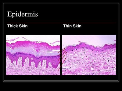
Thin vs. Thick Skin
Thick skin (palms, soles) has a prominent stratum lucidum and thicker stratum corneum. Thin skin covers most of the body and lacks stratum lucidum.
The Skeletal System: Osseous Tissue and Skeletal Structure
Bone Structure and Associated Features
Bones provide support, protection, and facilitate movement. Understanding bone anatomy is essential for identifying skeletal features.
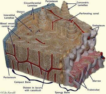
Periosteum: Outer fibrous layer covering bone.
Endosteum: Inner lining of bone cavities.
Compact Bone: Dense, strong bone tissue.
Spongy Bone: Porous, lightweight bone tissue.
Medullary Cavity: Central cavity containing bone marrow.
Lacuna: Small cavity housing osteocyte.
Osteocyte: Mature bone cell.
Central (Haversian) Canal: Contains blood vessels and nerves.
Canaliculi: Tiny channels connecting osteocytes.
Concentric Lamellae: Rings of bone matrix around central canal.
Circumferential Lamellae: Outer rings beneath periosteum.
Interstitial Lamellae: Fill spaces between osteons.
Osteon: Structural unit of compact bone.
Long Bone Anatomy
Key Features of Long Bones
Long bones have distinct regions and structures important for growth and function.
Diaphysis: Shaft of the bone.
Epiphysis: Ends of the bone.
Epiphyseal Line/Plate: Site of bone growth.
Medullary Cavity: Contains marrow.
Compact Bone Tissue: Dense outer layer.
Spongy Bone Tissue: Inner, porous layer.
Periosteum: Outer covering.
Endosteum: Inner lining.
Nutrient Foramen: Opening for blood vessels.
Joints & Ligaments
Types and Structures
Joints connect bones and allow movement. Ligaments stabilize joints.
Synovial Joint: Freely movable, contains synovial fluid.
Cartilaginous Joint: Slightly movable, connected by cartilage.
Fibrous Joint: Immovable, connected by fibrous tissue.
Bursa: Fluid-filled sac reducing friction.
Articular Cartilage: Covers bone surfaces in joints.
Knee Joint
Anterior Cruciate Ligament (ACL): Prevents forward movement of tibia.
Posterior Cruciate Ligament (PCL): Prevents backward movement of tibia.
Tibial (Medial) Collateral Ligament: Stabilizes inner knee.
Fibular (Lateral) Collateral Ligament: Stabilizes outer knee.
Patellar Ligament: Connects patella to tibia.
Medial/Lateral Meniscus: Shock absorbers.
Shoulder Joint
Acromioclavicular Ligament: Connects acromion to clavicle.
Coracoacromial Ligament: Connects coracoid to acromion.
Coracohumeral Ligament: Connects coracoid to humerus.
Coracoclavicular Ligament: Connects coracoid to clavicle.
Transverse Humeral Ligament: Holds biceps tendon in place.
Bones for Exam 1
Axial and Appendicular Skeleton
The skeleton is divided into axial (skull, vertebrae, thoracic cage) and appendicular (limbs, girdles) regions. Identification of bones and their features is essential.
Skull Bones: Frontal, parietal, temporal, occipital, sphenoid, ethmoid, nasal, maxilla, mandible, zygomatic, vomer, palatine, lacrimal.
Misc Bones: Hyoid.
Vertebrae: Cervical (7), Atlas (C1), Axis (C2), Thoracic (12), Lumbar (5), Sacrum, Coccyx.
Thoracic Cage: Sternum (manubrium, body), ribs (true, false, floating).
Pectoral Girdle & Upper Limbs: Scapula, clavicle, humerus, ulna, radius, carpals, metacarpals, phalanges.
Pelvic Girdle & Lower Limbs: Os Coxa, femur, tibia, fibula, patella, tarsals, metatarsals, phalanges.
Example: The femur is the longest bone in the body and is part of the appendicular skeleton.
Bone Type | Examples |
|---|---|
Axial Skeleton | Skull, vertebrae, thoracic cage |
Appendicular Skeleton | Limbs, girdles |
Additional info: The study guide covers foundational concepts in anatomy and physiology, including cell structure, tissue types, skin, bone, and joints, directly relevant to ANP college course chapters 1-8.
