 Back
BIOL 71 Lab Practical Study Guide: Foundations of Anatomy, Cells, Tissues, and Histology
Study Guide - Smart Notes
 Tailored notes based on your materials, expanded with key definitions, examples, and context.
Cell Biology
Cell Structure and Organelles
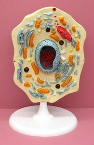
The cell is the basic structural and functional unit of life. Eukaryotic cells contain various organelles, each with specialized functions essential for cellular processes.
Plasma Membrane: The boundary of the cell, regulates entry and exit of substances.
Microvilli: Finger-like projections that increase surface area for absorption.
Cilia: Hair-like structures for movement of substances across cell surfaces.
Flagella: Tail-like structures for cell movement.
Cytoplasm: The region between the plasma membrane and nucleus, containing cytosol and organelles.
Nucleus: Contains genetic material (DNA); controls cell activities.
Nucleolus: Site of ribosome synthesis within the nucleus.
Nuclear Pores: Openings in the nuclear envelope for transport of molecules.
Chromatin: DNA and protein complex; condenses to form chromosomes during cell division.
Endoplasmic Reticulum (ER): Rough ER (with ribosomes) synthesizes proteins; Smooth ER synthesizes lipids.
Ribosome: Site of protein synthesis.
Golgi Apparatus: Modifies, sorts, and packages proteins and lipids for secretion or internal use.
Cytoskeleton: Network of protein filaments for cell shape, movement, and division.
Secretory Vesicle: Transports substances to the cell membrane for release.
Peroxisome: Breaks down fatty acids and detoxifies harmful substances.
Lysosome: Contains digestive enzymes for breakdown of waste.
Mitochondria: Powerhouse of the cell; site of ATP production.
Microscopy
Microscope Parts and Functions
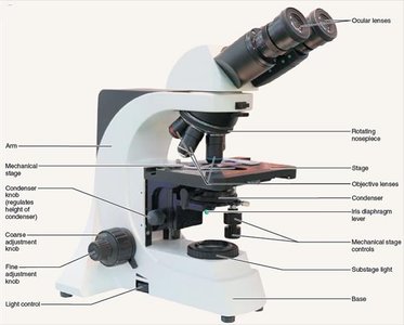
Microscopes are essential tools for studying cells and tissues. Understanding their parts and functions is crucial for laboratory work.
Ocular lens: Eyepiece for viewing specimens.
Objective lens: Provides various magnifications (4X, 10X, 40X, 100X).
Stage: Platform where slides are placed.
Coarse/Fine adjustment: Focuses the specimen.
Condenser: Focuses light onto the specimen.
Light control: Adjusts illumination.
Total magnification: Product of ocular and objective lens magnifications.
Field of view: Area visible through the microscope.
Resolution: Ability to distinguish two close points as separate.
Tissue Types
Overview of Major Tissue Types
Tissues are groups of cells with similar structure and function. The four primary tissue types are epithelial, connective, muscle, and nervous tissue.
Epithelial Tissue: Covers surfaces, lines cavities, forms glands.
Connective Tissue: Supports, protects, and binds other tissues. Includes blood, bone, cartilage, areolar, dense, and adipose tissues.
Muscle Tissue: Responsible for movement; includes skeletal, cardiac, and smooth muscle.
Nervous Tissue: Conducts electrical impulses; found in brain, spinal cord, and nerves.
Blood Tissue
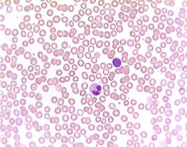
Blood is a connective tissue composed of cells suspended in plasma. It transports oxygen, nutrients, and waste.
Erythrocytes: Red blood cells, carry oxygen.
Leukocytes: White blood cells, defend against pathogens.
Platelets: Aid in blood clotting.
Bone Tissue
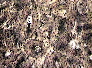
Bone is a rigid connective tissue providing structural support and protection. It contains osteocytes in lacunae, surrounded by a matrix of collagen and minerals.
Osteon: Structural unit of compact bone.
Central (Haversian) canal: Contains blood vessels and nerves.
Lacuna: Small cavity housing an osteocyte.
Lamellae: Layers of bone matrix.
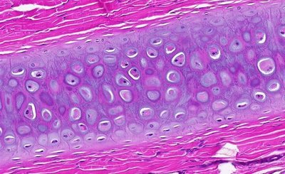
Cartilage Tissue
Cartilage is a flexible connective tissue found in joints, ear, nose, and respiratory tract. Chondrocytes reside in lacunae within a gel-like matrix.
Hyaline cartilage: Most common type, provides support and flexibility.
Chondrocyte: Cartilage cell within a lacuna.
Extracellular matrix: Gel-like substance surrounding cells.
Adipose Tissue
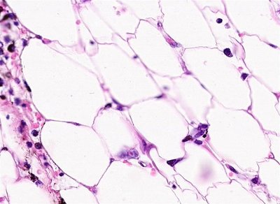
Adipose tissue is a type of connective tissue specialized for fat storage. It insulates and protects organs.
Adipocyte: Fat cell storing triglycerides.
Function: Energy storage, insulation, cushioning.
Muscle Tissue Types
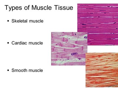
Muscle tissue is classified into three types based on structure and function.
Skeletal Muscle: Voluntary, striated, attached to bones.
Cardiac Muscle: Involuntary, striated, found in heart, contains intercalated discs.
Smooth Muscle: Involuntary, non-striated, found in walls of organs.
Epithelial Tissues
Classification and Structure
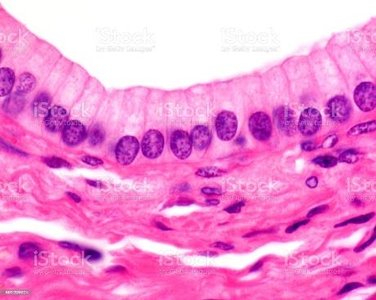
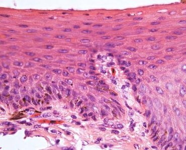
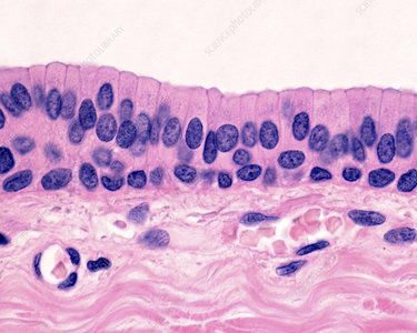
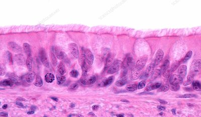
Epithelial tissues are classified by cell shape and number of layers. They line surfaces and cavities, and form glands.
Simple Squamous: Single layer of flat cells; allows diffusion.
Simple Cuboidal: Single layer of cube-shaped cells; absorption and secretion.
Simple Columnar: Single layer of tall cells; absorption and secretion.
Ciliated Pseudostratified Columnar: Appears layered, but all cells touch basement membrane; cilia move mucus.
Stratified Squamous: Multiple layers; protection against abrasion.
Stratified Cuboidal/Columnar: Multiple layers; rare, found in ducts.
Associated Structures: Cilia, apical/basal surfaces, basement membrane, lumen.
Skin Histology and Accessory Structures
Layers and Structures of the Skin
The skin is composed of multiple layers and accessory structures, providing protection, sensation, and thermoregulation.
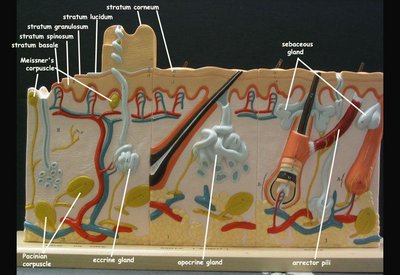
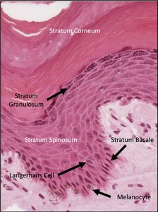
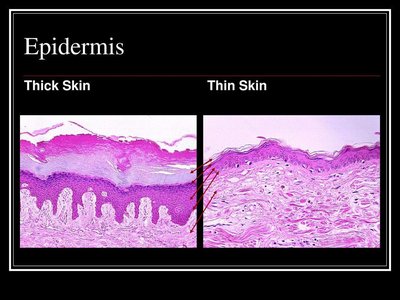
Epidermis: Outermost layer; includes stratum basale, spinosum, granulosum, lucidum, corneum.
Dermis: Middle layer; papillary and reticular regions, contains blood vessels and nerves.
Hypodermis: Subcutaneous layer; adipose and connective tissue.
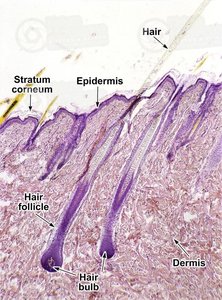
Accessory Structures: Hair shaft, follicle, arrector pili muscle, sebaceous gland, sweat gland.
Bone Structure
Compact and Spongy Bone
Bones are composed of compact and spongy tissue, each with distinct structures and functions.
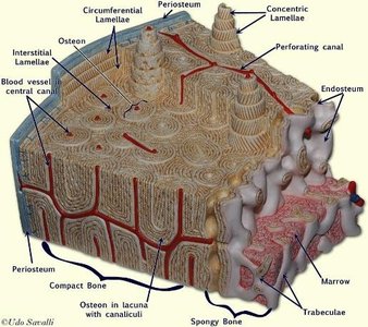
Periosteum: Outer fibrous layer covering bone.
Endosteum: Inner lining of medullary cavity and trabeculae.
Compact Bone: Dense, organized in osteons.
Spongy Bone: Porous, contains trabeculae and marrow.
Medullary Cavity: Central cavity containing bone marrow.
Lacunae vs. Osteocytes: Lacunae are spaces; osteocytes are cells within them.
Additional info:
These notes cover foundational concepts from Ch. 1-5 (cell biology, microscopy, tissues, skin, bone) and are directly relevant to the ANP college course. Images included are only those that clearly reinforce the explanation of the adjacent paragraph.
