 Back
BackBlood and the Cardiovascular System: Structure, Function, and Components
Study Guide - Smart Notes
 Tailored notes based on your materials, expanded with key definitions, examples, and context.
Blood and the Cardiovascular System
Overview of the Cardiovascular System
The cardiovascular system is essential for transporting substances throughout the body and maintaining homeostasis. It consists of the heart (a pump), blood vessels (conducting hoses), and blood (a fluid connective tissue).
Components and Functions of Blood
Major Functions of Blood
Transport: Carries dissolved gases (O2, CO2), nutrients, hormones, and metabolic wastes.
Regulation: Maintains pH, ion composition, and body temperature.
Protection: Restricts fluid loss at injury sites, defends against toxins and pathogens.
Stabilization: Stabilizes body temperature and interstitial fluid composition.
Physical Characteristics of Blood
Temperature: 38ºC (100.4ºF)
Viscosity: High (thicker than water)
pH: Slightly alkaline (7.35–7.45)
Volume: ~7% of body weight (e.g., 5.25 L in a 75-kg adult)
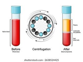
Composition of Blood
Whole Blood: Consists of plasma and formed elements.
Plasma: Fluid matrix (~55% of blood volume), mostly water (92%), with proteins and solutes.
Formed Elements: Red blood cells (RBCs), white blood cells (WBCs), and platelets.
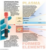
Plasma Proteins
Albumins (60%): Maintain osmotic pressure, transport fatty acids and hormones.
Globulins (35%): Include antibodies (immunoglobulins) and transport proteins.
Fibrinogen (4%): Functions in blood clotting; converted to fibrin.
Other Proteins (1%): Enzymes and hormones.
Formed Elements
Red Blood Cells (Erythrocytes): Transport oxygen and carbon dioxide.
White Blood Cells (Leukocytes): Defend against pathogens.
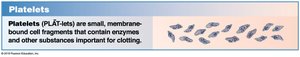
Platelets (Thrombocytes): Involved in clotting.
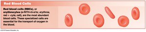
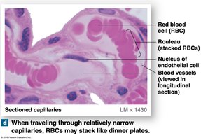
Red Blood Cells (RBCs)
Structure and Function
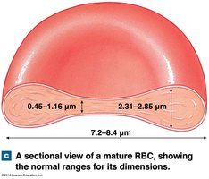
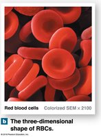
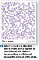
RBCs are small, biconcave discs that lack nuclei and most organelles. Their shape increases surface area for gas exchange and allows flexibility in capillaries.
Count: 4.5–6.3 million/μL (males), 4.2–5.5 million/μL (females)
Hematocrit: Percentage of blood volume occupied by RBCs (46% in males, 42% in females)
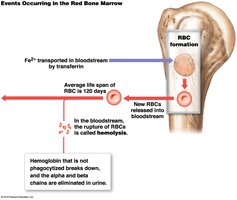
Lifespan: ~120 days
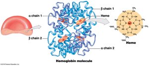
Hemoglobin (Hb)
Hemoglobin is a protein in RBCs responsible for transporting oxygen and carbon dioxide. Each molecule consists of four globular subunits, each with a heme group containing iron.
Normal Hb: 14–18 g/dL (males), 12–16 g/dL (females)
Oxyhemoglobin (HbO2): Formed when iron binds oxygen.
Deoxyhemoglobin: Hemoglobin without oxygen.
Fetal Hemoglobin: Binds oxygen more readily than adult hemoglobin.
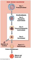
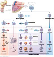
Red Blood Cell Production (Erythropoiesis)
Erythropoiesis is the process of RBC formation, occurring in red bone marrow. It is regulated by the hormone erythropoietin (EPO), secreted by the kidneys and liver in response to hypoxia.
Stages: Myeloid stem cell → Proerythroblast → Erythroblast stages → Reticulocyte → Mature RBC
Requirements: Amino acids, iron, folic acid, vitamins B12 and B6
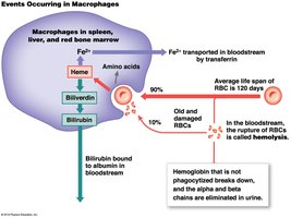
Hemoglobin and Iron Recycling
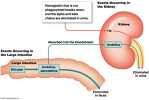
Old or damaged RBCs are engulfed by macrophages in the spleen, liver, and bone marrow. Hemoglobin is broken down, and iron is recycled for new RBC production. Biliverdin and bilirubin are produced from heme breakdown and excreted in bile.

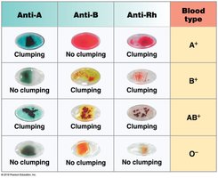
Blood Types
ABO and Rh Blood Groups
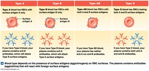
Blood type is determined by the presence or absence of specific surface antigens (agglutinogens) on RBCs: A, B, and Rh (D). The four main blood types are A, B, AB, and O. The Rh group determines if blood is positive or negative.
Type A: Surface antigen A, anti-B antibodies
Type B: Surface antigen B, anti-A antibodies
Type AB: Both antigens, no anti-A or anti-B antibodies
Type O: No antigens, both anti-A and anti-B antibodies
Rh+: Rh antigen present
Rh−: Rh antigen absent
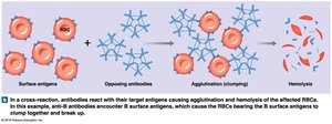
Transfusion Reactions and Compatibility
If incompatible blood is transfused, antibodies in the recipient's plasma react with donor RBC antigens, causing agglutination and hemolysis (cross-reaction). Compatibility testing is essential before transfusions.
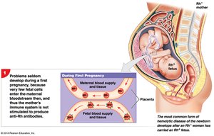
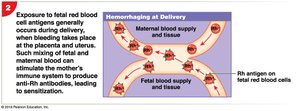
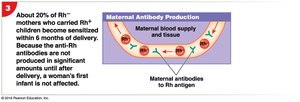
Hemolytic Disease of the Newborn (HDN)
HDN occurs when an Rh− mother carries an Rh+ fetus, leading to maternal antibody production against fetal RBCs in subsequent pregnancies.
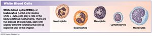
White Blood Cells (WBCs)
Types and Functions
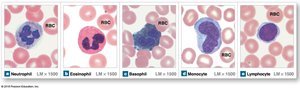
WBCs (leukocytes) are involved in defending the body against pathogens, removing toxins and wastes, and attacking abnormal or damaged cells. They are classified as granulocytes or agranulocytes.
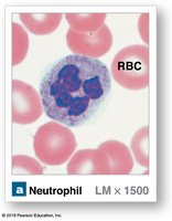
Neutrophils: Phagocytize bacteria, most abundant WBC.
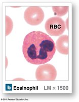
Eosinophils: Attack parasites, reduce inflammation.
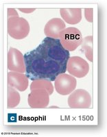
Basophils: Release histamine and heparin in damaged tissues.
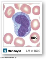
Monocytes: Become macrophages, engulf large pathogens.
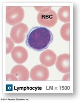
Lymphocytes: Specific immunity (T cells, B cells, NK cells).
WBC Production and Regulation
WBCs are produced in bone marrow from hemocytoblasts. Colony-stimulating factors (CSFs) regulate the production of specific WBC types.
Platelets
Structure and Function
Platelets are small, membrane-bound cell fragments essential for blood clotting. They circulate for 9–12 days and are removed by the spleen.
Release clotting chemicals
Form temporary plugs in vessel walls
Reduce size of vessel breaks
Hemostasis
Phases of Hemostasis
Hemostasis is the process of stopping blood loss after injury and involves three phases:
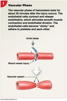
Vascular Phase: Vascular spasm constricts the vessel (immediate response).
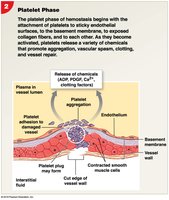
Platelet Phase: Platelets adhere to exposed surfaces and aggregate to form a plug (within 15 seconds).
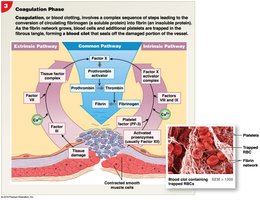
Coagulation Phase: Clotting factors activate to form a fibrin meshwork (within 30 seconds).
Clot Retraction and Fibrinolysis
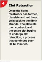
After clot formation, platelets contract to reduce the size of the break (clot retraction). Fibrinolysis is the process of dissolving the clot as healing occurs.
Summary Table: Main Components of Blood
Component | Percentage | Main Function |
|---|---|---|
Plasma | ~55% | Transport of nutrients, hormones, and waste; maintains osmotic balance |
Red Blood Cells | ~44% | Transport oxygen and carbon dioxide |
White Blood Cells | <1% | Defense against pathogens |
Platelets | <1% | Blood clotting |
Additional info: This guide covers the essential aspects of blood structure, function, and clinical relevance for ANP college students, including the mechanisms of hemostasis and the importance of blood typing in transfusion medicine.
