 Back
BackBlood and the Cardiovascular System: Structure, Function, and Disorders
Study Guide - Smart Notes
 Tailored notes based on your materials, expanded with key definitions, examples, and context.
Blood and the Circulatory System
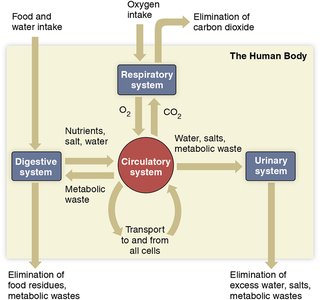
Overview of the Circulatory System
The circulatory system is essential for transporting materials throughout the body, maintaining homeostasis, and defending against disease. It consists of the heart, blood vessels, and blood. The system interacts closely with the respiratory, digestive, and urinary systems to exchange gases, nutrients, and wastes.
Primary function: Transport materials to and from all cells.
Other functions: Regulation of body temperature, water levels, pH, and defense against disease and injury.
Blood: A Specialized Connective Tissue
Blood is a specialized connective tissue composed of cells and cell fragments suspended in plasma. It originates from bone marrow and constitutes about 8% of body weight (5-6L in men, 4-5L in women).
Three crucial tasks:
Transportation of nutrients and wastes
Regulation of body temperature, water levels, and pH
Defense against pathogens and blood loss
Blood Composition
Formed Elements (45%)
Red blood cells (RBCs): Transport O2 to tissues and CO2 away from tissues.
White blood cells (WBCs): Defend the body against invaders and abnormal cells.
Platelets: Involved in blood clotting.
Plasma (55%)
Water: Main component of plasma.
Electrolytes: Na, K, Cl, HCO3, Ca, H, Mg – regulate cell function and excitability.
Proteins: Albumins (water balance), globulins (transport, immunity), clotting proteins.
Hormones: Chemical messengers.
Gases: O2 and CO2.
Nutrients & wastes: Glucose, urea, etc.

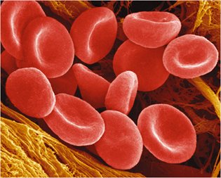
Red Blood Cells and Hemoglobin
Structure and Function of RBCs
Red blood cells, or erythrocytes, are highly specialized for gas transport. They are flexible, lack a nucleus and organelles, and are packed with hemoglobin.
Hemoglobin: Protein that binds O2 and CO2.
Each RBC contains about 300 million hemoglobin molecules.
RBCs are biconcave, increasing surface area for gas exchange.
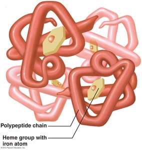
Hemoglobin and Oxygen Transport
Each hemoglobin molecule has 4 polypeptide chains, each with a heme group containing an iron atom.
O2 binds to iron in the heme group; one RBC can carry up to 1.2 billion O2 molecules.
Oxyhemoglobin: Hemoglobin bound to O2.
Deoxyhemoglobin: Hemoglobin without O2.
Hemoglobin also transports about 25% of CO2 (at a different site than O2).
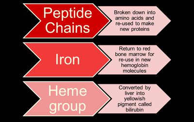
Recycling of Red Blood Cell Components
Old and damaged RBCs are removed by macrophages in the liver and spleen. Their components are recycled:
Component | Fate |
|---|---|
Peptide Chains | Broken down into amino acids for new proteins |
Iron | Returned to bone marrow for new hemoglobin |
Heme Group | Converted to bilirubin, excreted in bile |
Blood Cell Production and Regulation
Hematopoiesis: Formation of Blood Cells
All blood cells originate from stem cells in red bone marrow. These stem cells differentiate into various types of blood cells and platelets.
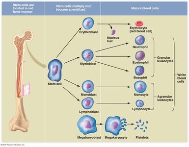
Erythroblasts: Immature RBCs that fill with hemoglobin and lose their nucleus to become erythrocytes.
Megakaryocytes: Large cells that fragment to form platelets.
Leukocytes: Differentiate into various types of WBCs (granular and agranular).
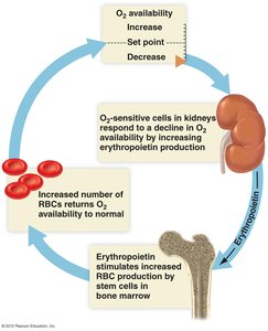
Regulation of RBC Production
RBC production is regulated by the hormone erythropoietin, released by the kidneys in response to low blood oxygen levels. This is a negative feedback mechanism.
White Blood Cells and Platelets
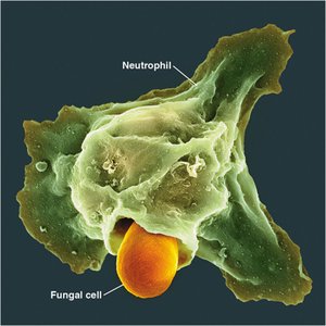
White Blood Cells (Leukocytes)
Granular leukocytes: Neutrophils, eosinophils, basophils (lifespan: hours to days).
Agranular leukocytes: Monocytes, lymphocytes (lifespan: days to years).
WBCs defend against pathogens, remove debris, and are involved in immune responses.
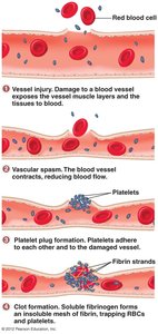
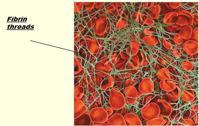
Platelets and Hemostasis
Platelets are cell fragments essential for blood clotting. Hemostasis involves three stages:
Vascular spasm (constriction of blood vessels)
Platelet plug formation
Blood clotting (coagulation)
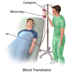
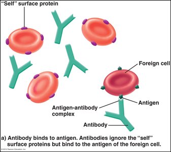
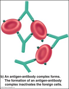
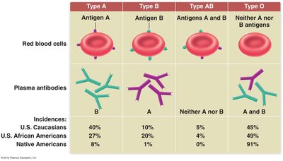
Blood Types and Transfusions
ABO Blood Group System
Blood types are determined by the presence of specific antigens on RBC surfaces. Antibodies in plasma react against foreign antigens, leading to agglutination if incompatible blood is transfused.
Blood Type | RBC Antigens | Plasma Antibodies |
|---|---|---|
A | A | Anti-B |
B | B | Anti-A |
AB | A and B | None |
O | None | Anti-A and Anti-B |
Rh Factor
The Rh antigen is another important surface protein. Rh-negative individuals can develop antibodies if exposed to Rh-positive blood, which is especially significant in pregnancy.
Blood Disorders
Anemia
Iron-deficiency anemia: Insufficient iron for hemoglobin synthesis.
Hemorrhagic anemia: Blood loss from injury or other causes.
Pernicious anemia: Vitamin B12 deficiency.
Hemolytic anemia: Destruction of RBCs (e.g., sickle-cell anemia).
Anemia due to renal failure: Reduced erythropoietin production.
Leukemia and Multiple Myeloma
Leukemia: Uncontrolled production of abnormal WBCs in bone marrow, crowding out normal cells.
Multiple myeloma: Cancer of plasma cells, leading to abnormal antibody production and weakened bones.
Thrombocytopenia
Reduction in platelet count, leading to increased bleeding and bruising.
Causes include infections, anemia, leukemia, drug reactions, etc.
Blood Vessels and the Heart
Types of Blood Vessels
Arteries: Thick-walled, carry blood away from the heart, withstand high pressure.
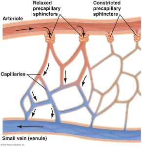
Arterioles: Smaller arteries, regulate blood flow to capillaries.
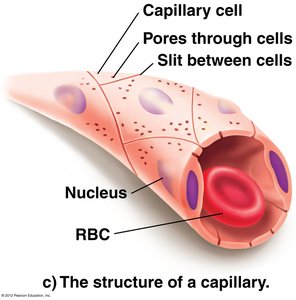
Capillaries: Microscopic vessels for exchange of gases, nutrients, and wastes.
Venules and veins: Return blood to the heart, have thinner walls and valves to prevent backflow.
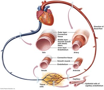

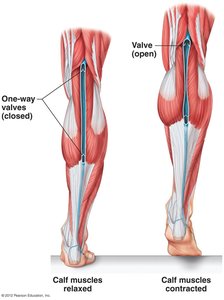
Venous Return Mechanisms
Skeletal muscle pump: Muscle contractions help push blood through veins.
One-way valves: Prevent backflow of blood.
Respiratory pump: Pressure changes during breathing assist venous return.
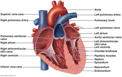
The Heart: Structure and Function
Four chambers: Right and left atria, right and left ventricles.
Valves: Atrioventricular (tricuspid, bicuspid/mitral) and semilunar (aortic, pulmonary) valves ensure one-way flow.
Pericardium: Protective outer layer.
Myocardium: Muscular middle layer responsible for contraction.
Endocardium: Inner endothelial layer.
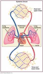
Pulmonary and Systemic Circuits
Pulmonary circuit: Right side of heart pumps deoxygenated blood to lungs for gas exchange.
Systemic circuit: Left side of heart pumps oxygenated blood to the body.
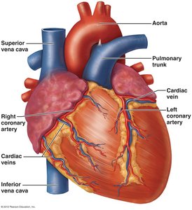
Coronary Circulation
The heart muscle (myocardium) receives its own blood supply via the coronary arteries. Cardiac veins return deoxygenated blood from the heart muscle to the right atrium.
Cardiac Cycle and Blood Pressure
The Cardiac Cycle
Systole: Contraction phase (atria then ventricles).
Diastole: Relaxation phase (atria and ventricles relax).
One complete cycle takes about 0.8 seconds.
Heart Sounds
"Lub": Closure of AV valves during ventricular systole.
"Dub": Closure of semilunar valves during ventricular diastole.
Heart murmurs: Abnormal sounds due to valve issues.
Electrical Conduction System
Sinoatrial (SA) node: Pacemaker, initiates heartbeat.
Atrioventricular (AV) node, AV bundle, Purkinje fibers: Coordinate contraction sequence.
Electrocardiogram (ECG): Records electrical activity of the heart (P wave, QRS complex, T wave).
Blood Pressure
Systolic pressure: Highest pressure during ventricular contraction.
Diastolic pressure: Lowest pressure during ventricular relaxation.
Normal BP: 120/80 mmHg or lower.
Cardiovascular Regulation and Disorders
Regulation of Blood Flow and Pressure
Cardiac output and arteriole diameter regulate arterial blood pressure.
Baroreceptors in arteries detect changes in pressure and initiate negative feedback to maintain homeostasis.
Cardiovascular Disorders
Angina: Chest pain due to reduced blood flow to the heart.
Heart attack (myocardial infarction): Death of heart tissue from prolonged oxygen deprivation.
Heart failure: Inefficient pumping leads to fluid buildup (congestive heart failure).
Embolism: Blockage of a blood vessel by a traveling clot or material.
Stroke: Interruption of blood supply to the brain (cerebrovascular accident).
Reducing Cardiovascular Risk
Non-modifiable risk factors: Sex, age, genetics.
Modifiable risk factors: Smoking, diet, exercise, blood pressure, diabetes control, stress management.
