 Back
Blood: Composition, Functions, and Clinical Significance
Study Guide - Smart Notes
 Tailored notes based on your materials, expanded with key definitions, examples, and context.
Blood Composition and Physical Properties
Overview of Blood
Blood is a specialized fluid connective tissue essential for transporting substances throughout the body. It consists of a liquid matrix called plasma and various formed elements (cells and cell fragments) suspended within it.
Plasma: The non-living, straw-colored fluid matrix, making up about 55% of blood volume.
Formed Elements: Living blood cells, including erythrocytes (red blood cells), leukocytes (white blood cells), and platelets (cell fragments).
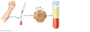
Blood Composition After Centrifugation
Plasma: ~55% of total blood volume; least dense component.
Buffy Coat: <1% of blood; contains leukocytes and platelets.
Erythrocytes: ~45% of blood (hematocrit); most dense component.
Hematocrit is the percentage of blood volume occupied by erythrocytes. Normal values: 47% ± 5% for males, 42% ± 5% for females.
Physical Characteristics and Volume
Sticky, opaque fluid with a metallic taste.
Color varies with oxygen content: scarlet (high O2), dark red (low O2).
pH: 7.35–7.45 (slightly alkaline).
Accounts for ~8% of body weight.
Average volume: 5–6 L (males), 4–5 L (females).
Functions of Blood
Distribution
Delivers oxygen and nutrients to body cells.
Transports metabolic wastes to lungs and kidneys for elimination.
Carries hormones from endocrine organs to target tissues.
Regulation
Maintains body temperature by absorbing and distributing heat.
Maintains normal pH using buffers (e.g., bicarbonate ions).
Maintains adequate fluid volume in the circulatory system.
Blood Plasma
Composition of Plasma
90% water.
Over 100 dissolved solutes: nutrients, gases, hormones, wastes, proteins, inorganic ions.
Plasma proteins (most abundant solutes): 60% albumin, 36% globulins, 4% fibrinogen.
Most plasma proteins are produced by the liver and remain in the blood.
Formed Elements of Blood
Erythrocytes (Red Blood Cells)
Erythrocytes are specialized for oxygen transport. Their structure is closely related to their function.
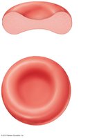
Biconcave disc shape increases surface area for gas exchange.
Over 97% hemoglobin (excluding water).
No mitochondria; ATP production is anaerobic, so they do not consume the oxygen they carry.
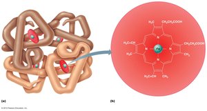
Hemoglobin Structure and Function
Hemoglobin is the protein responsible for oxygen transport in erythrocytes.
Composed of four polypeptide chains (two alpha, two beta) and four heme groups.
Each heme group contains an iron atom that binds one O2 molecule.
Each hemoglobin molecule can carry up to four oxygen molecules.
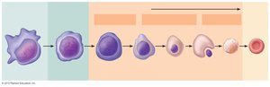
Hematopoiesis and Erythropoiesis
Hematopoiesis is the process of blood cell formation, occurring in red bone marrow. Erythropoiesis is the specific formation of red blood cells.
Occurs in axial skeleton, girdles, and proximal epiphyses of humerus and femur in adults.
Stages: Hematopoietic stem cell (hemocytoblast) → proerythroblast → erythroblast stages (basophilic, polychromatic, orthochromatic) → reticulocyte → erythrocyte.
Leukocytes (White Blood Cells)
General Features
Make up less than 1% of total blood volume (4,800–10,800 WBCs/μl).
Defend the body against disease.
Can leave capillaries (diapedesis) and move through tissues by ameboid motion and chemotaxis.
Leukocytosis: WBC count above 11,000/mm3, a normal response to infection.
Types of Leukocytes
Granulocytes: Neutrophils, eosinophils, basophils (contain visible cytoplasmic granules).
Agranulocytes: Lymphocytes, monocytes (lack visible granules).
Type | Relative Percentage |
|---|---|
Neutrophils | 50–70% |
Lymphocytes | 25–45% |
Monocytes | 3–8% |
Eosinophils | 2–4% |
Basophils | 0.5–1% |
Neutrophils
Most numerous WBC; multilobed nucleus.
Granules contain hydrolytic enzymes or defensins.
Very phagocytic; "bacteria slayers."
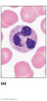
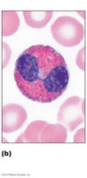
Eosinophils
Bilobed nucleus; red-staining granules.
Digest parasitic worms; involved in allergies and asthma.
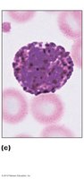
Basophils
Rarest WBC; large, purplish-black granules contain histamine.
Histamine acts as a vasodilator and attracts other WBCs to sites of inflammation.
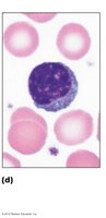
Lymphocytes
Second most numerous WBC; large, dark nucleus with thin rim of cytoplasm.
Crucial to adaptive immunity; mostly found in lymphoid tissues.
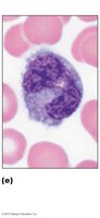
Monocytes
Largest leukocytes; kidney-shaped nucleus.
Differentiate into macrophages in tissues; phagocytic and activate lymphocytes.
Leukopoiesis
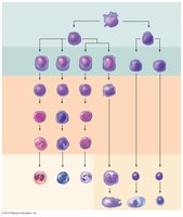
Leukopoiesis is the production of white blood cells, stimulated by interleukins and colony-stimulating factors (CSFs). All leukocytes originate from hematopoietic stem cells (hemocytoblasts).
Platelets
Structure and Function
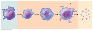
Cytoplasmic fragments of megakaryocytes.
Contain granules with serotonin, Ca2+, enzymes, ADP, and platelet-derived growth factor (PDGF).
Essential for blood clotting; form temporary platelet plugs to seal vessel breaks.
Normal count: 150,000–400,000/μl blood.
Platelet Formation
Regulated by thrombopoietin.
Megakaryoblast undergoes mitosis without cytokinesis, forming a large, multilobed megakaryocyte, which fragments into platelets.
Hemostasis
Overview
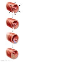
Hemostasis is the process of stopping bleeding, involving a rapid series of reactions that require clotting factors and substances released by platelets and injured tissues.
Step 1: Vascular Spasm – Vasoconstriction of damaged blood vessel.
Step 2: Platelet Plug Formation – Platelets adhere to exposed collagen fibers, become activated, and aggregate.
Step 3: Coagulation – Fibrin forms a mesh that traps blood cells, forming a clot.
Coagulation Pathways
Three phases: formation of prothrombin activator, conversion of prothrombin to thrombin, and formation of fibrin mesh.
Intrinsic and extrinsic pathways lead to the activation of prothrombin activator.
Vitamin K is required for the synthesis of several clotting factors.
Key Equations:
Clot Structure
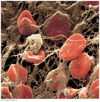
The final blood clot consists of erythrocytes trapped in a mesh of insoluble fibrin.
Human Blood Groups
Blood Group Antigens
RBC membranes have glycoprotein antigens (agglutinogens) that can trigger immune responses if foreign.
ABO and Rh antigens are clinically significant for transfusions.
ABO Blood Groups
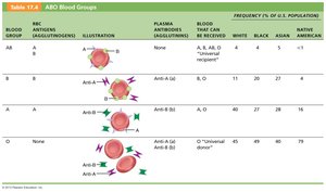
Four types: A, B, AB, O, based on presence/absence of A and B antigens.
Plasma contains preformed antibodies (agglutinins) against absent antigens.
Transfusion reactions occur if mismatched blood is given.
Blood Group | RBC Antigens | Plasma Antibodies | Blood Received |
|---|---|---|---|
A | A | Anti-B | A, O |
B | B | Anti-A | B, O |
AB | A, B | None | A, B, AB, O (universal recipient) |
O | None | Anti-A, Anti-B | O (universal donor) |
Blood Typing
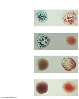
Blood typing involves mixing blood with anti-A and anti-B sera to observe agglutination, which indicates the presence of specific antigens.
Rh Blood Groups
52 named Rh antigens; C, D, and E are most common.
Rh+ indicates presence of D antigen (85% of Americans are Rh+).
