 Back
Blood Flow, Capillary Exchange, and Circulatory Pathways
Study Guide - Smart Notes
 Tailored notes based on your materials, expanded with key definitions, examples, and context.
Control of Blood Flow
Intrinsic and Extrinsic Control Mechanisms
Blood flow to tissues is regulated by both intrinsic (local) and extrinsic (systemic) mechanisms to ensure that each organ receives the appropriate amount of blood for its function.
Intrinsic controls (Autoregulation): Regulation originates within the tissue or organ itself, using local chemical signals (paracrines) or properties of muscle tissue. This allows organs to adjust their own blood flow by modifying arteriole diameter.
Extrinsic controls: Regulation comes from outside the tissue or organ, primarily via the sympathetic nervous system and hormones. These controls redistribute blood flow to where it is most needed and help maintain mean arterial pressure (MAP).
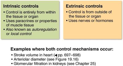
Examples: Both mechanisms influence stroke volume in the heart, arteriolar diameter, and glomerular filtration in the kidneys.
Redistribution of Blood Flow During Exercise
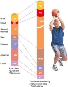
During exercise, blood flow is redistributed to meet the increased metabolic demands of skeletal muscle. Intrinsic controls cause vasodilation in active muscles, while extrinsic controls reduce blood flow to less active organs.
At rest, skeletal muscles receive about 20% of total blood flow; during strenuous exercise, this can exceed 70%.
MAP is maintained despite large changes in local blood flow.
Mechanisms of Arteriolar Control
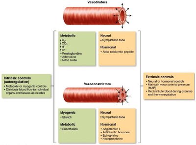
Arteriolar smooth muscle is regulated by a combination of metabolic, myogenic, neural, and hormonal factors. These mechanisms determine whether vasodilation or vasoconstriction occurs.
Vasodilators: Metabolic factors (e.g., low O2, high CO2, H+, adenosine, nitric oxide), neural (decreased sympathetic tone), and hormonal (atrial natriuretic peptide).
Vasoconstrictors: Myogenic (stretch), metabolic (endothelins), neural (increased sympathetic tone), and hormonal (angiotensin II, antidiuretic hormone, norepinephrine, epinephrine).
Autoregulation and Special Circulations
Autoregulation (Intrinsic Regulation)
Autoregulation ensures that blood flow matches the metabolic needs of tissues. It involves two main mechanisms:
Metabolic controls: Increased metabolic activity leads to decreased O2 and increased metabolic byproducts (H+, K+, adenosine, prostaglandins), causing vasodilation.
Myogenic controls: Vascular smooth muscle responds to changes in pressure. Increased stretch (high MAP) causes vasoconstriction; reduced stretch (low MAP) causes vasodilation.
Long-term autoregulation involves angiogenesis (formation of new blood vessels) in response to chronic changes in tissue demand.
Active Hyperemia in Skeletal Muscle
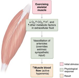
During muscle activity, local metabolic changes override sympathetic vasoconstriction, resulting in increased blood flow (active hyperemia).
Blood Flow in Special Areas
Brain: Maintains constant flow; sensitive to CO2 and pH changes.
Skin: Regulates temperature and acts as a blood reservoir; controlled by neural reflexes.
Lungs: Low O2 causes vasoconstriction, directing blood to well-ventilated areas.
Heart: Blood flow increases during diastole and with increased metabolic demand.
Capillary Exchange and Bulk Flow
Velocity of Blood Flow
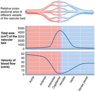
Blood flow velocity is fastest in the aorta and slowest in capillaries, allowing time for exchange of gases, nutrients, and wastes. Velocity is inversely related to the total cross-sectional area of the vessels.
Capillary Exchange Mechanisms
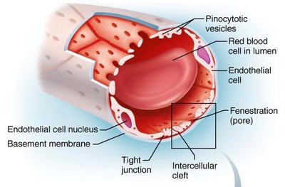
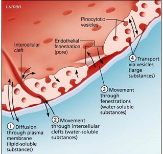
Substances move between blood and interstitial fluid via four main routes:
Direct diffusion through endothelial membranes (lipid-soluble molecules)
Passage through intercellular clefts (water-soluble solutes)
Passage through fenestrations (water-soluble solutes)
Active transport via pinocytotic vesicles or caveolae (large molecules such as proteins)
Bulk Flow: Fluid Movements Across Capillary Walls
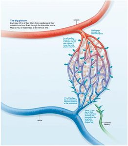
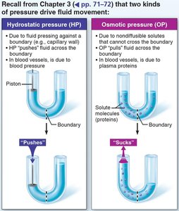
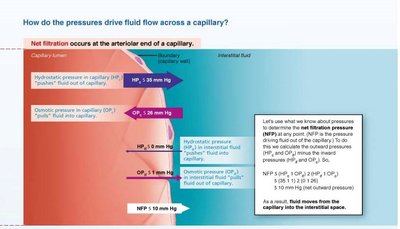
Bulk flow refers to the movement of fluid across capillary walls, driven by hydrostatic and osmotic pressures. This process determines the distribution of fluid between plasma and interstitial fluid.
Hydrostatic pressure (HP): The force exerted by fluid pressing against a wall; pushes fluid out of capillaries.
Osmotic pressure (OP): The force exerted by nondiffusible solutes (mainly plasma proteins); pulls fluid into capillaries.
Net Filtration Pressure (NFP)
The direction and amount of fluid movement are determined by the net filtration pressure:
At the arterial end, net filtration (fluid leaves capillary).
At the venous end, net reabsorption (fluid enters capillary).
Excess fluid is returned to the blood via the lymphatic system.
Clinical Correlation: Edema
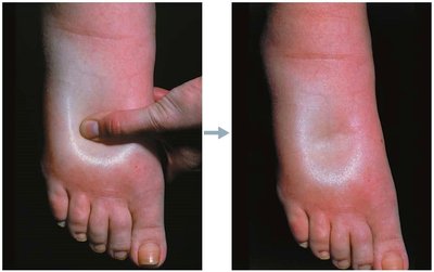
Edema is an abnormal accumulation of interstitial fluid, caused by increased capillary hydrostatic pressure, increased interstitial fluid osmotic pressure, decreased capillary colloid osmotic pressure, or impaired lymphatic drainage. It can impair tissue function by increasing the distance for diffusion of gases and nutrients.
Circulatory Pathways
Pulmonary and Systemic Circulation
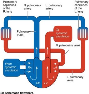
The vascular system consists of two main circuits:
Pulmonary circulation: Short loop from the heart to the lungs and back, for gas exchange.
Systemic circulation: Long loop from the heart to all body tissues and back, delivering oxygen and nutrients.
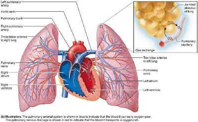
Systemic Circulation Overview
Systemic arteries run deep, while veins can be deep or superficial. Venous pathways are more interconnected and may have multiple names. The brain and digestive systems have unique venous drainage systems (dural sinuses and hepatic portal system, respectively).
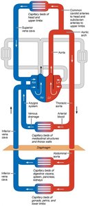
Major Arteries and Veins
Major arteries and veins are named according to the region, organ, or bone they serve. Arteries and veins often run side by side, but not always symmetrically. Diagrams use red for oxygen-rich blood and blue for oxygen-poor blood.
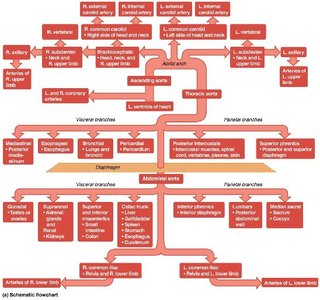
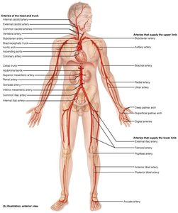
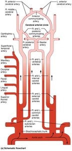
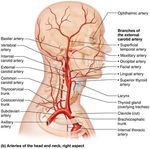
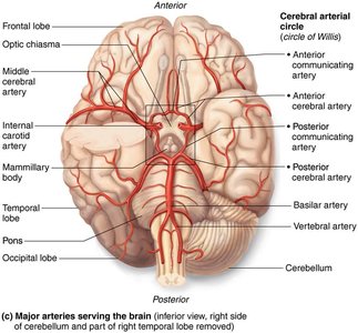
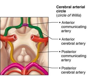
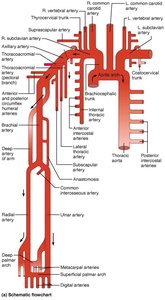
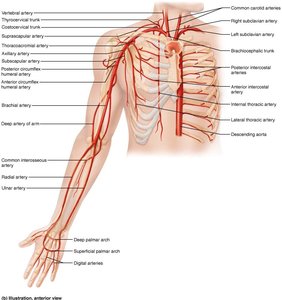
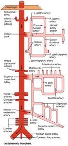
Additional info: For a complete understanding, students should refer to anatomical diagrams and practice tracing the flow of blood through both the pulmonary and systemic circuits, as well as identifying major vessels in the body.
