 Back
BackBlood Flow, Capillary Exchange, and Circulatory Pathways: ANP Study Guide
Study Guide - Smart Notes
 Tailored notes based on your materials, expanded with key definitions, examples, and context.
Control of Blood Flow
Intrinsic and Extrinsic Control Mechanisms
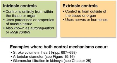
Blood flow to tissues is regulated by both intrinsic (local) and extrinsic (systemic) mechanisms. These controls ensure that each tissue receives the appropriate amount of blood for its metabolic needs.
Intrinsic controls: Originate within the tissue or organ itself. They use paracrines or properties of muscle tissue and are also known as autoregulation or local control.
Extrinsic controls: Originate from outside the tissue or organ, using nerves or hormones to regulate blood flow.
Examples: Both mechanisms influence stroke volume in the heart, arteriolar diameter, and glomerular filtration in the kidneys.
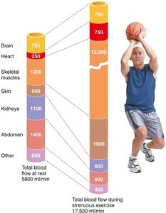
Redistribution of Blood Flow During Exercise
During strenuous exercise, blood flow is redistributed to meet the increased demands of skeletal muscles. Intrinsic controls dilate arterioles in active muscles, while extrinsic controls reduce flow to less active organs.
At rest: Skeletal muscles receive about 20% of total blood flow.
During exercise: Skeletal muscles can receive over 70% of blood flow, while flow to kidneys and digestive organs decreases.
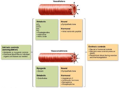
Mechanisms of Arteriolar Control
Arteriolar smooth muscle is regulated by both vasodilators and vasoconstrictors, which are influenced by intrinsic and extrinsic factors.
Vasodilators: Include metabolic factors (↓O2, ↑CO2, ↑H+, prostaglandins, adenosine, nitric oxide) and neural/hormonal factors (↓sympathetic tone, atrial natriuretic peptide).
Vasoconstrictors: Include myogenic stretch, endothelins, increased sympathetic tone, and hormones (angiotensin II, antidiuretic hormone, norepinephrine, epinephrine).
Blood Flow in Special Areas
Skeletal Muscle Blood Flow
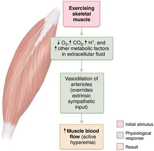
Blood flow to skeletal muscle increases during activity due to local metabolic changes, a process called active hyperemia.
Initial stimulus: Exercise increases metabolic factors in extracellular fluid.
Physiological response: Vasodilation of arterioles overrides extrinsic sympathetic input.
Result: Increased muscle blood flow.
Capillary Exchange
Velocity of Blood Flow
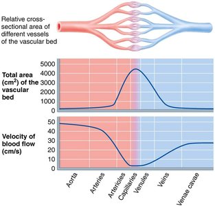
The velocity of blood flow changes throughout the circulatory system. It is fastest in the aorta and slowest in capillaries, allowing time for exchange of substances.
Speed is inversely related to total cross-sectional area: Capillaries have the largest area and slowest flow.
Capillary Transport Mechanisms
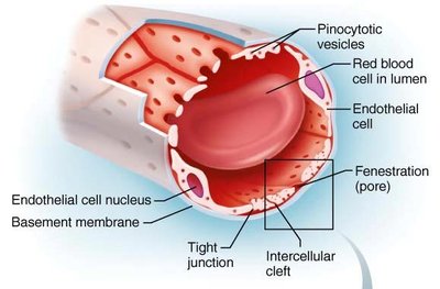
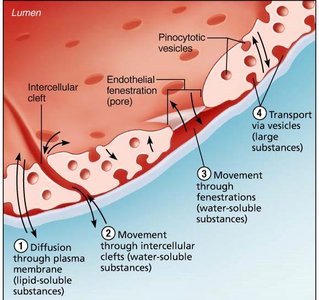
Capillaries allow exchange of gases, nutrients, and waste products between blood and tissues via four main routes:
Direct diffusion: Lipid-soluble molecules pass through endothelial membranes.
Intercellular clefts: Water-soluble solutes move through gaps between endothelial cells.
Fenestrations: Water-soluble solutes pass through pores in the endothelium.
Pinocytotic vesicles: Larger molecules are transported via vesicles.
Fluid Movements: Bulk Flow
Bulk Flow Across Capillary Walls
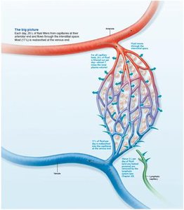
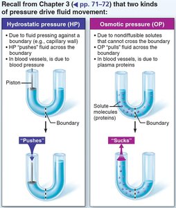
Bulk flow is the movement of fluid across capillary walls, driven by hydrostatic and osmotic pressures. It is essential for maintaining fluid balance between plasma and interstitial fluid.
Hydrostatic pressure (HP): The force exerted by fluid pressing against the capillary wall, pushing fluid out.
Osmotic pressure (OP): The force exerted by plasma proteins, pulling fluid into the capillary.
Hydrostatic and Osmotic Pressure Interactions
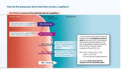
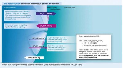
The direction and amount of fluid movement depend on the balance between hydrostatic and osmotic pressures. Net filtration pressure (NFP) determines whether fluid leaves or enters the capillary.
Equation:
At arterial end: Net fluid flow out (filtration).
At venous end: Net fluid flow in (reabsorption).
Excess fluid: Returned to blood via lymphatic system.
Clinical Homeostatic Imbalance: Edema
Causes and Effects of Edema
Edema is an abnormal increase in interstitial fluid, caused by imbalances in hydrostatic or osmotic pressures, or impaired lymphatic drainage.
Increased capillary hydrostatic pressure: Accelerates fluid loss from blood.
Increased interstitial fluid osmotic pressure: Inflammation increases permeability, allowing proteins to leak and pull fluid into interstitial space.
Decreased capillary colloid osmotic pressure: Hinders fluid return to blood, often due to hypoproteinemia.
Impaired lymphatic drainage: Blockage or removal of lymphatic vessels causes fluid accumulation.
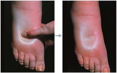
Pitting edema: Excess interstitial fluid in subcutaneous tissues causes visible indentation.
Circulatory Pathways
Pulmonary and Systemic Circulation
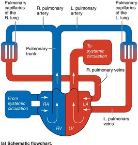
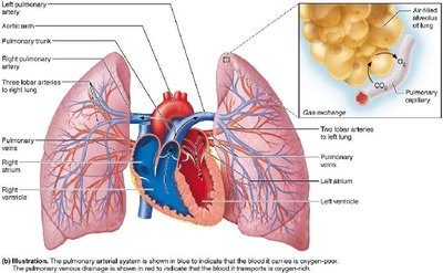
The vascular system consists of two main circulations: pulmonary (heart to lungs and back) and systemic (heart to body and back).
Pulmonary circulation: Short loop, low pressure, oxygen-poor blood travels from right ventricle to lungs and returns oxygen-rich blood to left atrium.
Systemic circulation: Long loop, high pressure, oxygen-rich blood travels from left ventricle to body and returns oxygen-poor blood to right atrium.
Overview of Systemic Circulation
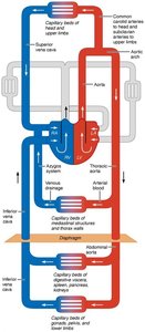
Systemic arteries and veins have distinct pathways and naming conventions. Arteries are deep, veins can be deep or superficial, and venous pathways are more interconnected.
Brain and digestive systems: Have unique venous drainage systems (dural venous sinuses, hepatic portal system).
Principal Vessels of the Systemic Circulation
Major Arteries
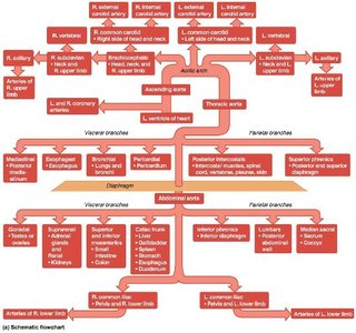
Arteries are named based on the region, organ, or bone they serve. They tend to run side by side with veins and nerves.
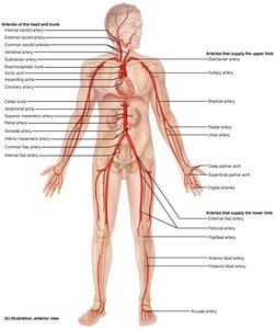
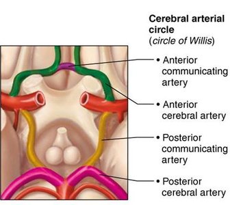
Arteries of the Head, Neck, and Brain
Arteries in these regions are bilaterally symmetrical, with some exceptions in the trunk.
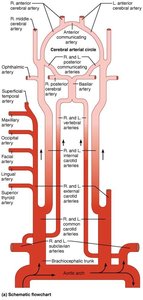
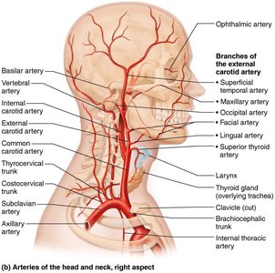
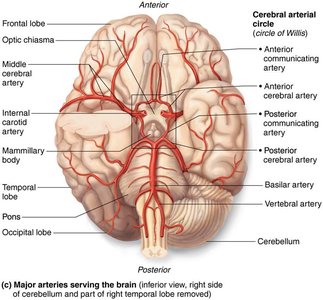
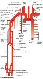
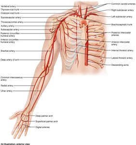
Arteries of the Right Upper Limb and Thorax
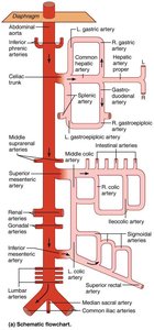
Arteries of the Abdomen
Arteries of the Right Pelvis and Lower Limb
Major Veins of the Systemic Circulation
Venous Drainage of the Head, Neck, and Brain
Veins of the Thorax and Right Upper Limb
Veins of the Abdomen
Veins of the Right Lower Limb
Developmental Aspects of Blood Vessels
Formation and Aging
Blood vessels develop from mesodermal cells forming blood islands, guided by vascular endothelial growth factor. Fetal shunts bypass nonfunctional organs, and vessel formation continues throughout life for growth, healing, and repair.
Congenital vascular problems: Rare.
Aging: May lead to varicose veins, atherosclerosis, and increased blood pressure.
