 Back
BackBlood Flow, Capillary Exchange, and Venous Circulation: Study Notes for Human Anatomy & Physiology
Study Guide - Smart Notes
 Tailored notes based on your materials, expanded with key definitions, examples, and context.
The Cardiovascular System: Blood Vessels
Key Concepts in Blood Flow Regulation
Blood flow through tissues is tightly regulated by both intrinsic (local) and extrinsic (systemic) mechanisms. These controls ensure that tissues receive adequate oxygen and nutrients while removing metabolic wastes.
Intrinsic controls (autoregulation): Local adjustments based on the immediate needs of individual organs.
Extrinsic controls: Systemic regulation to maintain mean arterial pressure (MAP) and direct blood flow where it is most needed.
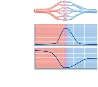
Slow blood flow through capillaries promotes efficient exchange of gases, nutrients, and bulk flow of fluids.
Functions of Tissue Perfusion
Tissue perfusion refers to the blood flow through body tissues, serving several essential functions:
Delivery of O2 and nutrients to tissue cells and removal of CO2 and wastes
Gas exchange in the lungs
Absorption of nutrients from the digestive tract
Formation of urine in the kidneys
Control of Blood Flow
Intrinsic (Local) Control
Intrinsic control, or autoregulation, is the regulation of blood flow from within the tissue or organ itself, independent of neural or hormonal input.
Local chemical factors (metabolic controls) and myogenic mechanisms adjust arteriole diameter.
Ensures that each organ receives blood flow according to its current needs.
Examples: Increased metabolic activity leads to vasodilation and increased blood flow.
Extrinsic Control
Extrinsic control involves regulation from outside the tissue or organ, primarily through the nervous system and hormones.
Maintains systemic blood pressure (MAP) and redistributes blood flow during exercise or thermoregulation.
Sympathetic nerves and hormones (e.g., epinephrine, norepinephrine, angiotensin II) act on arteriolar smooth muscle.
Comparison of Intrinsic and Extrinsic Controls
Intrinsic Controls | Extrinsic Controls |
|---|---|
Local chemicals (paracrines), muscle properties | Nerves (sympathetic), hormones |
Autoregulation/local control | Systemic regulation of MAP |
Distributes blood flow to organs as needed | Redistributes blood flow during exercise, stress |
Distribution of Blood Flow at Rest and During Exercise
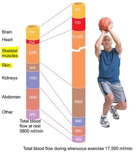
Blood flow is dynamically redistributed during physical activity to meet the metabolic demands of tissues.
At rest, most blood flows to abdominal organs, kidneys, and brain.
During strenuous exercise, blood flow increases dramatically to skeletal muscles and skin, while decreasing to the abdomen and kidneys.
Mechanisms of Arteriolar Control
Vasodilators: Nitric oxide (NO), prostaglandins, adenosine, decreased O2, increased CO2, H+, K+
Vasoconstrictors: Endothelins, sympathetic tone, angiotensin II, antidiuretic hormone
Long-Term Autoregulation
When short-term autoregulation is insufficient, long-term mechanisms such as angiogenesis (formation of new blood vessels) occur, especially in response to chronic low oxygen (e.g., high altitude, coronary artery blockage).
Blood Flow in Specific Organs
Skeletal Muscle
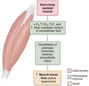
Blood flow to skeletal muscle is highly variable and increases significantly during exercise due to local metabolic controls overriding sympathetic vasoconstriction.
At rest: Myogenic and neural mechanisms predominate.
During exercise: Increased metabolic activity (active hyperemia) leads to vasodilation and increased blood flow.
Brain
The brain receives a constant blood supply (~750 ml/min) and is highly sensitive to ischemia. Cerebral blood flow is regulated by both metabolic and myogenic mechanisms.
Decreased pH or increased CO2 causes vasodilation.
Changes in MAP alter vessel diameter to maintain constant flow.
Skin
Blood flow to the skin helps regulate body temperature, ranging from 50 ml/min to 2500 ml/min.
Increased body temperature: Vasodilation allows heat loss.
Decreased body temperature: Vasoconstriction conserves heat.
Lungs
Pulmonary circulation exhibits unique autoregulatory responses:
Low O2 levels cause vasoconstriction (opposite of systemic circulation).
High O2 levels promote vasodilation, optimizing gas exchange.
Heart
Coronary blood flow is about 250 ml/min at rest and is regulated by myogenic mechanisms and local vasodilators (e.g., adenosine) during exercise.
Blood Flow Velocity and Capillary Exchange
Blood Flow Velocity
Blood flow velocity is inversely related to the total cross-sectional area of blood vessels. It is slowest in the capillaries, allowing time for exchange of materials.
Capillary Transport Mechanisms
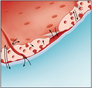
Capillaries allow exchange of substances between blood and tissues via several mechanisms:
Diffusion through plasma membrane (lipid-soluble substances)
Movement through intercellular clefts and fenestrations (water-soluble substances)
Transport via vesicles or caveolae (large substances)
Bulk Flow Across Capillary Walls
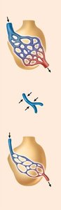
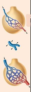
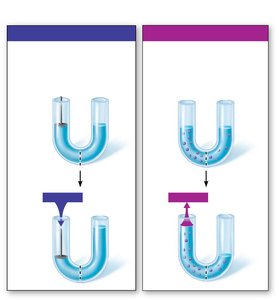
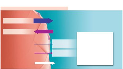
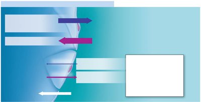
Bulk flow refers to the movement of fluid across capillary walls, driven by hydrostatic and osmotic pressures. This process maintains the interstitial environment and balances fluid between plasma and interstitial fluid.
Hydrostatic pressure (HP): Pushes fluid out of capillaries.
Osmotic pressure (OP): Pulls fluid into capillaries, mainly due to plasma proteins.
Fluid Movement in Capillary Beds
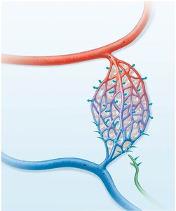
Each day, about 20 L of fluid is filtered out of capillaries, with 17 L reabsorbed and 3 L returned via the lymphatic system.
Net Filtration Pressure (NFP)
NFP determines the direction of fluid movement:
At the arteriolar end: Net outward pressure (filtration)
At the venous end: Net inward pressure (reabsorption)
Formula for NFP:
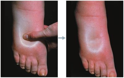
Homeostatic Imbalances: Edema
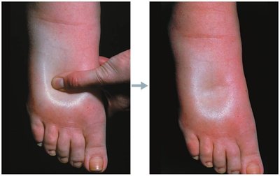
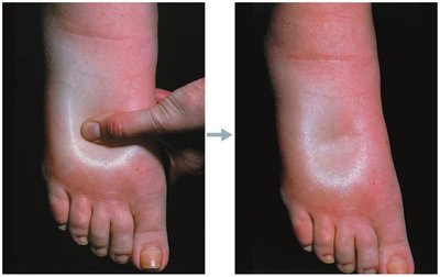
Edema is an abnormal accumulation of interstitial fluid, resulting from:
Increased capillary hydrostatic pressure (e.g., heart failure, venous blockage)
Increased interstitial fluid osmotic pressure (e.g., inflammation, anaphylaxis)
Decreased capillary colloid osmotic pressure (e.g., hypoproteinemia from malnutrition, liver disease)
Decreased lymphatic drainage (e.g., elephantiasis)
Major Veins of the Systemic Circulation
Overview of Major Veins
The systemic veins return deoxygenated blood from the body to the heart. The superior vena cava drains regions above the diaphragm, while the inferior vena cava drains regions below.
Major veins include the jugular, subclavian, brachiocephalic, renal, hepatic, iliac, and saphenous veins.
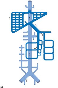
The hepatic portal system carries nutrient-rich blood from digestive organs to the liver.
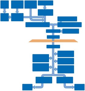
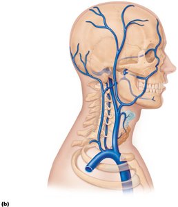
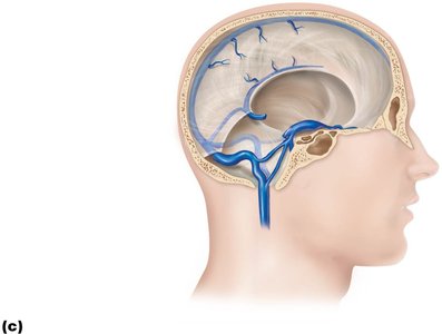
Venous Drainage of the Head, Neck, and Brain
Venous blood from the head and neck is collected by the internal and external jugular veins, vertebral veins, and dural venous sinuses.
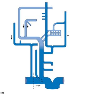
Veins of the Thorax and Upper Limb
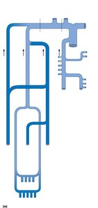
Venous return from the thorax and upper limb involves the subclavian, axillary, brachial, cephalic, basilic, and median cubital veins.
Veins of the Abdomen
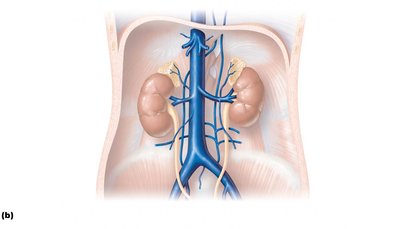
Abdominal veins include the inferior vena cava, renal, hepatic, gonadal, and lumbar veins. The hepatic portal system is a unique venous network connecting digestive organs to the liver.
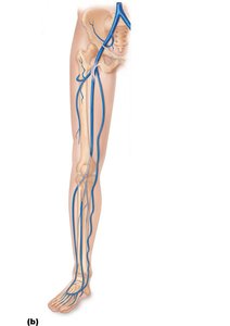
Veins of the Lower Limb
Venous drainage of the lower limb includes the femoral, great saphenous (the longest vein in the body), small saphenous, popliteal, tibial, and fibular veins.
Additional info: The hepatic portal system is essential for processing nutrients and detoxifying substances absorbed from the digestive tract before they enter the systemic circulation.
