 Back
BackBlood Pressure, Capillary Exchange, and Fetal Circulation
Study Guide - Smart Notes
 Tailored notes based on your materials, expanded with key definitions, examples, and context.
Blood Pressure and Its Measurement
Arterial Pressure Fluctuations
Blood pressure is the force exerted by circulating blood on the walls of blood vessels. It fluctuates with each heartbeat and is measured as two values:
Systolic Pressure: The peak pressure during ventricular contraction.
Diastolic Pressure: The minimum pressure at the end of ventricular relaxation.
Blood pressure is recorded as systolic over diastolic (e.g., 120/80 mm Hg).
Korotkoff sounds are the sounds heard when measuring blood pressure with a stethoscope and sphygmomanometer.
Pulse: The rhythmic, alternating changes in arterial pressure that can be felt at certain points on the body.
Pulse Pressure
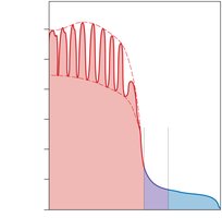
Pulse pressure is the difference between systolic and diastolic pressures. It reflects the force that the heart generates each time it contracts.
Pulse pressure diminishes over distance from the heart and is eliminated at the capillary level.
This is due to arterial recoil (elastic rebound) during diastole, which helps maintain blood flow.
Pulses can be felt at pressure points where arteries are close to the skin.
Venous Pressure
Venous pressure is much lower than arterial pressure. Two main mechanisms help blood return to the heart, especially from the lower body:
Muscular Compression: Skeletal muscles compress veins, pushing blood toward the heart.
Venous Valves: Prevent backflow of blood.
Respiratory Pump: Changes in thoracic pressure during breathing assist venous return.
Capillary Exchange
Capillary Pressures
Capillary pressure is the force exerted by blood within capillaries. It drops from about 35 mm Hg at the arterial end to 18 mm Hg at the venous end.
Capillaries are permeable to ions, nutrients, wastes, gases, and water.
Capillary pressure causes filtration of water and solutes out of the bloodstream into tissues.
Some materials are reabsorbed into capillaries; the remainder is picked up by lymphatic vessels.
Functions of Capillary Exchange
Capillary exchange is essential for maintaining homeostasis and tissue health. Its four main functions are:
Maintaining constant communication between plasma and interstitial fluid.
Speeding distribution of nutrients, hormones, and gases.
Assisting in the transport of insoluble molecules.
Flushing bacterial toxins and other chemicals to lymphatic tissues for immune defense.
Mechanisms of Capillary Exchange
Three primary mechanisms facilitate the movement of substances across capillary walls:
Diffusion: Movement of ions or molecules from high to low concentration.
Filtration: Movement of solute due to the "push" of water or hydrostatic pressure down fluid pressure gradients. Water is filtered out of the capillary by hydrostatic pressure.
Reabsorption: Water is reabsorbed into the capillary by osmosis, driven by osmotic pressure.
Forces Acting Across Capillary Walls
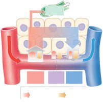
The movement of fluid across capillary walls is determined by the balance between capillary hydrostatic pressure (CHP) and blood osmotic pressure (BOP):
At the arterial end, CHP > BOP, so fluid is forced out (filtration).
At the midpoint, CHP = BOP, so there is no net movement.
At the venous end, BOP > CHP, so fluid moves back in (reabsorption).
Excess fluid (~3.6 L/day) enters lymphatic vessels and returns to circulation.
Fetal Circulation
Unique Features of Fetal Circulation
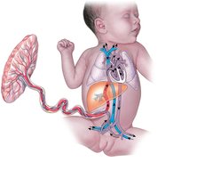
Fetal circulation includes several structures that allow blood to bypass the nonfunctional fetal lungs and efficiently exchange gases and nutrients with the placenta:
Foramen ovale: An opening between the right and left atria, allowing blood to bypass the fetal lungs.
Ductus arteriosus: A vessel connecting the pulmonary trunk to the aorta, further bypassing the lungs.
Ductus venosus: Allows oxygenated blood from the umbilical vein to bypass the liver and flow directly into the inferior vena cava.
Umbilical arteries and vein: Carry deoxygenated blood from the fetus to the placenta and oxygenated blood from the placenta to the fetus, respectively.
Blood Flow in the Fetus
Oxygenated blood returns from the placenta via the umbilical vein.
Blood bypasses the liver through the ductus venosus and enters the inferior vena cava.
Blood flows through the foramen ovale and ductus arteriosus to bypass the lungs.
Deoxygenated blood returns to the placenta via the umbilical arteries.
Checkpoint Questions
Factors contributing to total peripheral resistance: Vessel diameter, blood viscosity, vessel length, and turbulence.
Blood pressure is greater at the aorta than at the inferior vena cava because pressure decreases as blood moves through the systemic circuit due to resistance.
Fainting in the heat: Standing in the hot sun causes vasodilation and pooling of blood in the lower extremities, reducing venous return and cerebral perfusion, leading to fainting.
