 Back
BackBlood: Structure, Function, and Clinical Relevance
Study Guide - Smart Notes
 Tailored notes based on your materials, expanded with key definitions, examples, and context.
Blood: Structure, Function, and Clinical Relevance
Cardiovascular System Overview
The cardiovascular system is the primary transport system in the human body, consisting of the heart and blood vessels (arteries, veins, and capillaries). Blood flows through these vessels, delivering essential substances and removing waste products.
Transportation: Oxygen, nutrients, hormones, and waste products.
Regulation: pH, body temperature, and fluid volume.
Protection: Clot formation and defense against infection.

Composition of Blood
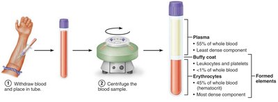
Blood is the only fluid tissue in the body, composed of plasma and formed elements. It is more dense and viscous than water, with a pH of 7.35–7.45 and a temperature of approximately 38°C. Blood constitutes about 8% of body weight.
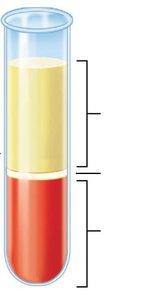
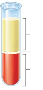
Plasma: The liquid component, making up 55% of whole blood.
Formed Elements: Erythrocytes (RBCs), leukocytes (WBCs), and platelets.
Blood Plasma
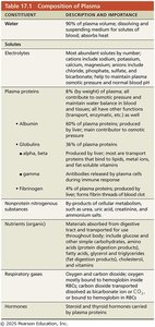
Plasma is the straw-colored, liquid portion of blood, consisting mainly of water and dissolved substances. It is crucial for maintaining osmotic pressure and transporting nutrients, hormones, and waste products.
Water: 90% of plasma volume.
Solutes: Electrolytes, plasma proteins (albumin, globulins, fibrinogen), nutrients, respiratory gases, hormones.
Albumin: Most abundant plasma protein, major contributor to osmotic pressure.
Constituent | Description and Importance |
|---|---|
Water | 90% of plasma volume; dissolving and suspending medium for solutes; absorbs heat |
Electrolytes | Maintain osmotic balance, pH, and membrane potential |
Plasma proteins | Albumin (osmotic pressure), globulins (transport, immunity), fibrinogen (clotting) |
Nutrients | Glucose, amino acids, fatty acids, vitamins |
Respiratory gases | O2 and CO2 |
Hormones | Regulate physiological processes |
Formed Elements of Blood
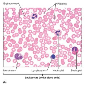
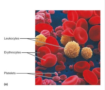
Formed elements are the cellular components of blood, including erythrocytes, leukocytes, and platelets. Most are short-lived and replenished by stem cells in the bone marrow.
Erythrocytes (RBCs): 45% of whole blood; transport oxygen and carbon dioxide.
Leukocytes (WBCs): <1% of whole blood; defense against disease.
Platelets: <1% of whole blood; essential for clotting.
Cell Type | Description | Cells/μL | Life Span | Function |
|---|---|---|---|---|
Erythrocytes | Biconcave, anucleate | 4-6 million | 100-120 days | Transport O2 and CO2 |
Leukocytes | Complete cells | 4,800-10,800 | Varies | Defense |
Platelets | Cell fragments | 150,000-400,000 | 5-10 days | Clotting |
Leukocytes: Types and Functions
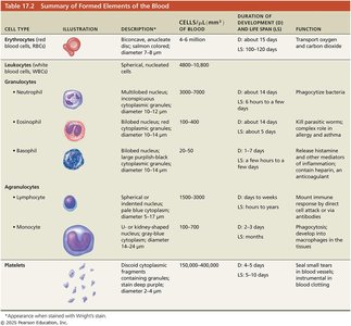
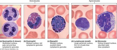
Leukocytes are the only complete cells among formed elements and are crucial for immune defense. They are classified as granulocytes (neutrophils, eosinophils, basophils) and agranulocytes (lymphocytes, monocytes).
Neutrophils: Phagocytize bacteria.
Lymphocytes: T cells (attack virus-infected cells), B cells (produce antibodies).
Monocytes: Differentiate into macrophages, phagocytic.
Eosinophils: Kill parasitic worms, modulate allergic responses.
Basophils: Release histamine, mediate inflammation.
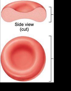
Erythrocytes: Structure and Function
Erythrocytes (RBCs) are small, biconcave, anucleate cells specialized for gas transport. Their shape increases surface area for efficient gas exchange, and they contain hemoglobin, the oxygen-carrying protein.
Biconcave shape: Maximizes surface area for gas exchange.
No mitochondria: ATP production is anaerobic, preventing O2 consumption.
Hemoglobin: 97% of cell volume (excluding water).
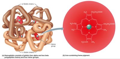
Hemoglobin Structure and Function
Hemoglobin is a protein composed of four globin chains (two alpha, two beta) and four heme groups, each containing an iron ion that binds oxygen. Hemoglobin also transports carbon dioxide as carbaminohemoglobin.
Oxyhemoglobin: Oxygen-bound form, bright red.
Deoxyhemoglobin: Oxygen released, dark red.
Carbaminohemoglobin: CO2 bound to globin.
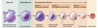
Hematopoiesis and Erythropoiesis
Hematopoiesis is the formation of all blood cells from hematopoietic stem cells in red bone marrow. Erythropoiesis is the specific process of RBC formation, regulated by erythropoietin (EPO) produced by the kidneys in response to hypoxia.
Stem cell: Hemocytoblast.
Committed cell: Proerythroblast.
Developmental pathway: Basophilic erythroblast → polychromatic erythroblast → orthochromatic erythroblast → reticulocyte → erythrocyte.
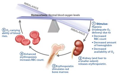
Dietary Requirements and RBC Life Cycle
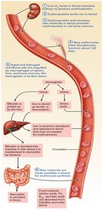
RBC production requires amino acids, lipids, carbohydrates, iron, vitamin B12, and folic acid. Iron is stored as ferritin and hemosiderin, transported by transferrin. RBCs have a lifespan of 100–120 days and are removed by the spleen.
Hemoglobin breakdown: Heme → bilirubin (excreted as stercobilin), iron recycled.
Globin: Amino acids recycled.
Blood Disorders
Anemia
Anemia is characterized by abnormally low oxygen-carrying capacity due to decreased RBC production, increased RBC destruction, or blood loss. Symptoms include fatigue, pallor, shortness of breath, and chills.
Iron deficiency anemia: Insufficient iron for hemoglobin synthesis.
Pernicious anemia: Vitamin B12 deficiency.
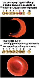
Hemolytic anemia: RBC destruction (e.g., sickle-cell anemia, thalassemia).
Polycythemia
Polycythemia is an abnormal excess of RBCs, increasing blood viscosity and causing sluggish blood flow. It may result from bone marrow cancer (polycythemia vera) or conditions stimulating EPO production (secondary polycythemia).

Platelets and Hemostasis
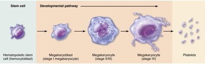
Platelets are cytoplasmic fragments of megakaryocytes, essential for clotting. They degenerate in about 10 days and are produced in bone marrow and lungs.
Normal count: 150,000–400,000 platelets/μL.
Contain: Serotonin, Ca2+, enzymes, ADP, PDGF.
Hemostasis: Steps and Clotting Factors
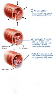
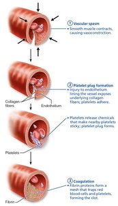
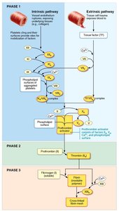
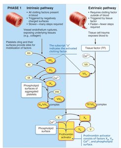
Hemostasis is the process of stopping bleeding, involving three principal steps: vascular spasm, platelet plug formation, and coagulation. Platelets and clotting factors play critical roles in each step.
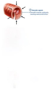
Vascular spasm: Vasoconstriction in response to vessel damage.
Platelet plug formation: Platelets adhere to exposed collagen, become activated, and aggregate.
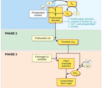
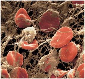
Coagulation: Reinforces plug with fibrin mesh, transforming blood from liquid to gel.
Factor Number | Factor Name | Source | Function |
|---|---|---|---|
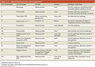
I | Fibrinogen | Liver | Converted to fibrin |
II | Prothrombin | Liver | Converted to thrombin |
III | Tissue factor | Tissue cells | Activates extrinsic pathway |
IV | Calcium ions | Plasma | Required for all stages |
V-XIII | Various | Liver, platelets | Clotting cascade |
Fibrinolytic System and Anticoagulants
The fibrinolytic system removes clots after repair. Plasminogen is converted to plasmin, which digests fibrin. Anticoagulants (e.g., antithrombin III, heparin) inhibit clotting to prevent excessive or inappropriate clot formation.
Clot retraction: Platelets contract, pulling vessel edges together.
Repair: PDGF and VEGF stimulate tissue regeneration.
Fibrinolysis: Plasmin digests fibrin, dissolving the clot.
Hemostasis Disorders
Disorders include thromboembolic conditions (thrombus, embolus), bleeding disorders (thrombocytopenia, hemophilia), and disseminated intravascular coagulation (DIC).
Thrombus: Clot in unbroken vessel.
Embolus: Freely floating clot.
Thrombocytopenia: Low platelet count.
Hemophilia: Hereditary deficiency of clotting factors.
DIC: Widespread clotting and bleeding.
Blood Types and Transfusion Reactions
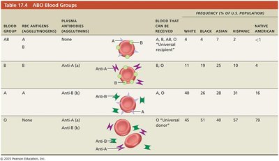
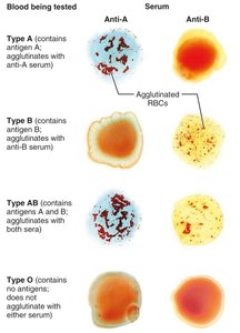
Blood types are determined by antigens (agglutinogens) on RBC membranes. The ABO and Rh systems are clinically significant. Transfusion of incompatible blood can cause agglutination and hemolysis, leading to severe reactions.
ABO system: Presence/absence of A and B antigens.
Rh system: Presence/absence of D antigen (Rh+ or Rh–).
Transfusion reactions: Recipient antibodies attack donor RBCs.
Blood Group | Antigens | Antibodies | Blood Received |
|---|---|---|---|
AB | A, B | None | A, B, AB, O |
A | A | Anti-B | A, O |
B | B | Anti-A | B, O |
O | None | Anti-A, Anti-B | O |
Rh Blood Groups and Hemolytic Disease of the Newborn
Rh incompatibility can cause hemolytic disease of the newborn (erythroblastosis fetalis) if an Rh– mother carries an Rh+ fetus. RhoGAM prevents sensitization.
Rh+: D antigen present.
Rh–: D antigen absent; anti-Rh antibodies form after exposure.
Developmental Aspects of Blood
Blood cells develop from mesenchymal blood islands in the embryo. Fetal blood cells form in the yolk sac, liver, and spleen, with hemoglobin F having higher O2 affinity than adult hemoglobin A. Blood diseases of aging include chronic leukemias, anemias, and clotting disorders.
