 Back
BackBlood: Structure, Function, and Clinical Relevance
Study Guide - Smart Notes
 Tailored notes based on your materials, expanded with key definitions, examples, and context.
Blood: Structure, Function, and Clinical Relevance
Overview and Functions of Blood
Blood is a specialized connective tissue essential for the transport of substances, protection against disease, and regulation of physiological parameters. It is composed of a liquid matrix (plasma) and formed elements (cells and cell fragments).
Transport: Delivers oxygen, nutrients, hormones, and removes metabolic wastes.
Protection: Prevents blood loss (clotting) and combats infection (immune cells).
Regulation: Maintains body temperature, pH (buffering systems), and fluid volume.
Physical Characteristics: Blood is slightly alkaline (pH 7.35–7.45), hot (38°C), sticky, salty, and ranges from scarlet (oxygen-rich) to dark red (oxygen-poor).
Blood Composition and Hematocrit
Blood consists of plasma (the liquid portion) and formed elements (cells and cell fragments). The percentage of blood volume occupied by red blood cells is called the hematocrit (47% ± 5% for males; 42% ± 5% for females).
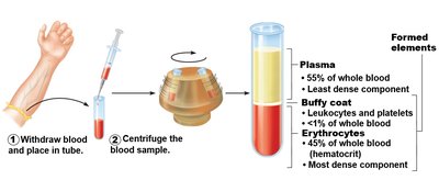

Plasma: The Fluid Matrix
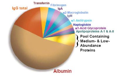
Plasma makes up about 55% of blood volume and is approximately 90% water. It contains dissolved solutes such as nutrients, gases, hormones, wastes, and ions. Plasma proteins (7–8%) are primarily produced by the liver and serve various functions:
Albumins: Most abundant; maintain osmotic pressure and transport substances.
Globulins: Include antibodies and transport proteins (alpha, beta, gamma types).
Fibrinogen: Precursor to fibrin, essential for blood clotting.
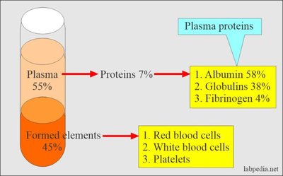
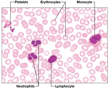
Formed Elements of Blood
The formed elements include erythrocytes (red blood cells), leukocytes (white blood cells), and platelets (cell fragments). Only leukocytes are complete cells; erythrocytes lack nuclei and organelles, and platelets are fragments of larger cells.
Erythrocytes: Transport oxygen and carbon dioxide.
Leukocytes: Defend against pathogens.
Platelets: Involved in clotting.
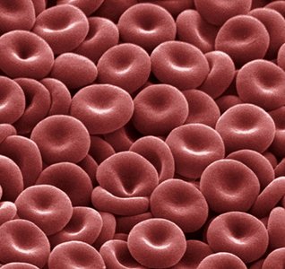
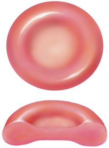
Erythrocytes (Red Blood Cells)
Structure and Function
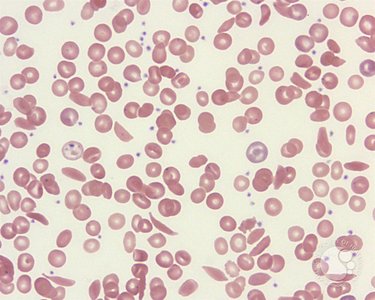
Erythrocytes are small (7.5 µm), biconcave discs that lack nuclei and organelles. Their shape increases surface area for gas exchange and allows flexibility to traverse capillaries. They generate ATP anaerobically, so they do not consume the oxygen they transport.
Main Functions:
Transport oxygen from lungs to tissues.
Carry some carbon dioxide from tissues to lungs.
Hemoglobin: Each erythrocyte contains about 250 million hemoglobin molecules, each capable of binding four oxygen molecules.
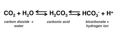
Gas Transport and Buffering
Hemoglobin binds oxygen reversibly and also assists in carbon dioxide transport. The majority of CO2 is transported as bicarbonate ions in plasma, following this reaction:
$\mathrm{CO_2 + H_2O \leftrightarrow H_2CO_3 \leftrightarrow HCO_3^- + H^+}$
Hematopoiesis and Erythropoiesis
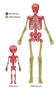
Blood cells are produced in the red bone marrow through a process called hematopoiesis. Erythrocyte production (erythropoiesis) is regulated by the hormone erythropoietin (EPO), primarily released by the kidneys in response to hypoxia (low oxygen levels).
Stages: Hematopoietic stem cell → Proerythroblast → Erythroblast → Reticulocyte → Erythrocyte
Regulation: Negative feedback ensures stable RBC count to avoid hypoxemia or increased blood viscosity.
Dietary Requirements for Erythropoiesis
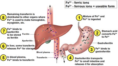
Essential nutrients include amino acids, lipids, carbohydrates, iron (stored as ferritin or transported by transferrin), and B-complex vitamins (B12 and folic acid) for DNA synthesis.
Erythrocyte Death and Disposal
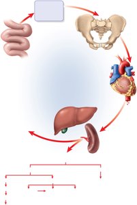
After about 120 days, erythrocytes are removed by the liver and spleen. Hemoglobin is broken down: globin is recycled as amino acids, iron is stored or reused, and heme is converted to bilirubin (excreted in bile).
Anemias
Anemia is a condition characterized by a reduced oxygen-carrying capacity of blood. It can result from blood loss, inadequate erythropoiesis, or increased erythrocyte destruction (hemolysis). Common types include iron-deficiency anemia, pernicious anemia (B12 deficiency), and hemolytic anemia (e.g., sickle cell disease).
Symptoms: Fatigue, pallor, shortness of breath, edema, and increased heart rate.
Blood Types and Transfusion Compatibility
ABO and Rh Blood Groups
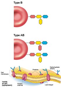
Blood types are determined by the presence or absence of specific antigens (agglutinogens) on erythrocyte membranes. The ABO system is based on A and B antigens; the Rh system is based on the D antigen.
Antigens: Unique molecules on RBC surfaces; recognized as self or foreign.
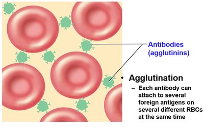
Antibodies (agglutinins): Proteins in plasma that bind foreign antigens, causing agglutination (clumping) and possible transfusion reactions.
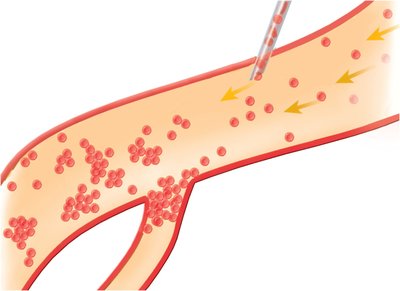
Transfusion Reactions
If incompatible blood is transfused, antibodies in the recipient's plasma bind to donor RBC antigens, causing agglutination and hemolysis. This can block blood vessels and cause severe complications.
ABO Blood Types
Blood Type | Antigens on RBCs | Antibodies in Plasma | Transfusion Compatibility |
|---|---|---|---|
A | A | Anti-B | Can receive A, O |
B | B | Anti-A | Can receive B, O |
AB | A and B | None | Universal recipient |
O | None | Anti-A, Anti-B | Universal donor |
Rh Factor
The Rh group is determined by the presence (Rh+) or absence (Rh-) of the D antigen. Anti-D antibodies are only produced in Rh- individuals after exposure to Rh+ blood (e.g., during pregnancy or transfusion).
Hemolytic Disease of the Newborn: Occurs if an Rh- mother carries an Rh+ fetus and produces anti-D antibodies that attack fetal RBCs in subsequent pregnancies.
Leukocytes (White Blood Cells)
General Function and Types
Leukocytes are complete cells that defend the body against pathogens. They are classified as granulocytes (neutrophils, eosinophils, basophils) and agranulocytes (lymphocytes, monocytes).
Neutrophils: Phagocytize bacteria; most abundant.
Eosinophils: Combat parasites and allergens.
Basophils: Release histamine and heparin; rare.
Lymphocytes: T cells (cell-mediated immunity), B cells (antibody production).
Monocytes: Differentiate into macrophages; phagocytize pathogens and debris.
Platelets and Hemostasis
Platelet Structure and Function
Platelets are cytoplasmic fragments derived from megakaryocytes. They play a crucial role in hemostasis (the control of bleeding) by forming plugs, releasing clotting factors, and facilitating vessel repair.
Hemostasis Steps:
Vascular spasm (vasoconstriction)
Platelet plug formation
Coagulation (fibrin mesh formation)
Clotting Disorders
Thromboembolic Disorders: Unwanted clot formation (thrombus, embolus).
Bleeding Disorders: Impaired clotting (e.g., hemophilia, vitamin K deficiency).
Summary Table: Major Blood Components
Component | Main Function | Key Features |
|---|---|---|
Plasma | Transport, osmotic balance | 90% water, proteins, nutrients, wastes |
Erythrocytes | O2 and CO2 transport | Biconcave, no nucleus, hemoglobin-rich |
Leukocytes | Immunity | Granulocytes & agranulocytes, nucleated |
Platelets | Clotting | Cell fragments, form plugs |
