 Back
BackBlood: Structure, Function, and Clinical Relevance
Study Guide - Smart Notes
 Tailored notes based on your materials, expanded with key definitions, examples, and context.
Blood: Structure, Function, and Clinical Relevance
Introduction to Blood
Blood is a specialized fluid connective tissue essential for transporting substances throughout the body. It consists of cells suspended in a liquid matrix called plasma, and plays a critical role in homeostasis, immunity, and hemostasis.
Composition of Whole Blood
Main Components
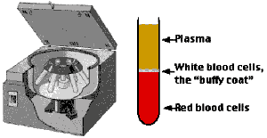
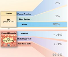
Plasma: The liquid matrix, making up 55% of blood volume, containing water, proteins, and dissolved solutes.
Formed Elements: Cellular components including red blood cells (RBCs), white blood cells (WBCs), and platelets.
Physical Characteristics of Blood
Temperature: 38°C (100.4°F), slightly higher than body temperature.
pH: Slightly alkaline (7.35–7.45).
Volume: ~7% of body weight (5–6 L in males, 4–5 L in females).
Plasma Composition
Water: 92% of plasma volume.
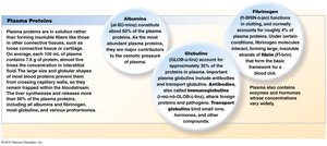
Plasma Proteins: 7% (Albumins, Globulins, Fibrinogen).
Other Solutes: 1% (electrolytes, nutrients, gases, waste products).
Plasma Proteins
Albumins (60%): Maintain osmotic pressure and transport substances.
Globulins (35%): Include antibodies (immunoglobulins) and transport proteins.
Fibrinogen (4%): Soluble protein involved in blood clotting; converts to fibrin during coagulation.
Formed Elements of Blood
Types of Formed Elements
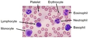
Red Blood Cells (Erythrocytes): Transport oxygen and carbon dioxide.
White Blood Cells (Leukocytes): Immune defense; five main types (neutrophils, eosinophils, basophils, lymphocytes, monocytes).
Platelets (Thrombocytes): Cell fragments involved in clotting.
Relative Abundance
RBCs: 99.9% of formed elements
WBCs and Platelets: <0.1% each
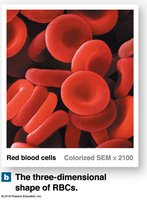
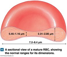
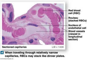
Red Blood Cells (RBCs)
Structure and Function
RBCs are biconcave discs specialized for efficient gas transport. Their shape increases surface area for gas exchange and allows flexibility to pass through capillaries. They lack nuclei and organelles, relying on anaerobic metabolism, and have a lifespan of ~120 days.
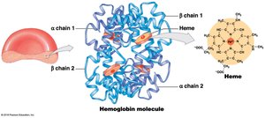
Hemoglobin
Structure: Each hemoglobin molecule has four subunits (2 alpha, 2 beta chains), each with a heme group containing iron (Fe) that binds oxygen.
Function: Transports O2 (as oxyhemoglobin) and CO2 (as carbaminohemoglobin). Releases O2 in tissues and binds CO2 for transport to the lungs.
Normal Hemoglobin Levels: Males: 14–18 g/dL; Females: 12–16 g/dL.
RBC Production (Erythropoiesis)
Occurs in red bone marrow from hematopoietic stem cells (HSCs).
Requires amino acids, iron, vitamins B12, B6, and folic acid.
Stimulated by erythropoietin (EPO), a hormone released by the kidneys in response to hypoxia.
RBC Disorders
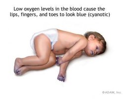
Anemia: Low RBC count, hematocrit, or hemoglobin; causes fatigue, dizziness, cyanosis.
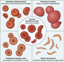
Sickle-cell anemia: Abnormal hemoglobin causes sickle-shaped RBCs.
Pernicious anemia: Vitamin B12 deficiency due to lack of intrinsic factor.
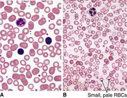
Iron deficiency anemia: Small, pale RBCs due to insufficient iron.
Polycythemia: Excess RBCs, increasing blood viscosity and risk of thrombosis.
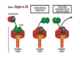
Carbon monoxide poisoning: CO binds hemoglobin with high affinity, preventing O2 transport.


Blood Types and Transfusion Compatibility
ABO and Rh Blood Groups
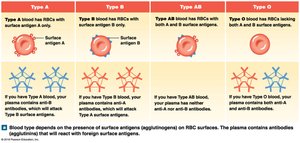
Blood types are determined by the presence or absence of antigens (A, B, Rh) on RBC membranes. The ABO system classifies blood as A, B, AB, or O, while the Rh system classifies as positive (+) or negative (–).
Universal donor: O–
Universal recipient: AB+
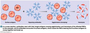
Transfusion Reactions (Cross-reactions)
If incompatible blood is transfused, antibodies in the recipient's plasma react with donor RBC antigens, causing agglutination and hemolysis.
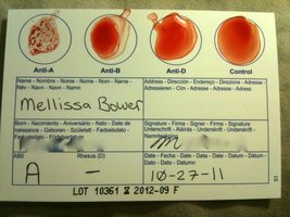
Blood Typing Tests
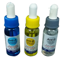
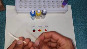
Blood samples are mixed with anti-A, anti-B, and anti-D (Rh) sera. Agglutination indicates the presence of the corresponding antigen.
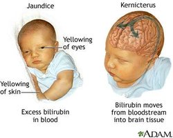
Hemolytic Disease of the Newborn (HDN)
Occurs when an Rh– mother carries an Rh+ fetus. Maternal anti-Rh antibodies can cross the placenta in subsequent pregnancies, destroying fetal RBCs. Prevention involves RhoGAM administration to the mother.
White Blood Cells (WBCs)
Types and Functions
WBCs (leukocytes) are immune cells with nuclei and organelles. They defend against pathogens, remove toxins, and attack abnormal cells. Normal count: 5,000–10,000/μL.
Neutrophils: Phagocytize bacteria; most abundant.
Eosinophils: Combat parasites and allergens.
Basophils: Release histamine; involved in inflammation.
Lymphocytes: Adaptive immunity (B and T cells).
Monocytes: Become macrophages in tissues.

WBC Production and Disorders
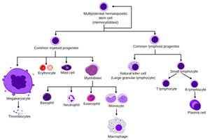
All blood cells originate from hemocytoblasts in bone marrow.
Myeloid stem cells produce all WBCs except lymphocytes; lymphoid stem cells produce lymphocytes.
Leukopenia: Low WBC count.
Leukemia: Excessive proliferation of abnormal WBCs.
Platelets and Hemostasis
Platelets
Cell fragments derived from megakaryocytes in bone marrow.
Essential for blood clotting; lifespan 9–12 days.
Normal count: 150,000–500,000/μL.
Hemostasis (Blood Clotting)
Hemostasis is the process of stopping bleeding, involving three phases:
Vascular Phase: Vasoconstriction and release of endothelins.
Platelet Phase: Platelet adhesion and aggregation to form a plug.
Coagulation Phase: Cascade of reactions converting fibrinogen to fibrin, forming a stable clot. Involves intrinsic, extrinsic, and common pathways.
Essential factors: Calcium ions, vitamin K, clotting factors I–XIII.
Thrombin catalyzes fibrin formation.
Anticoagulants (e.g., heparin, antithrombin) regulate clotting.
Fibrinolysis: Plasmin digests fibrin, dissolving the clot.
Platelet and Clotting Disorders
Thrombocytopenia: Low platelet count.
Thrombocytosis: High platelet count.
Thrombosis: Formation of a clot within a vessel (thrombus); if it moves, called an embolus.
Hemophilia: Genetic deficiency of clotting factors (e.g., Factor VIII), leading to impaired clotting.
Summary Table: Main Components of Blood
Component | Percentage of Whole Blood | Main Function |
|---|---|---|
Plasma | ~55% | Transport of nutrients, hormones, proteins, waste |
Red Blood Cells | ~45% | Oxygen and carbon dioxide transport |
White Blood Cells | <1% | Immune defense |
Platelets | <1% | Blood clotting |
Key Equations
Hematocrit (HCT):
Example Application
If a blood sample has a total volume of 5 mL and a plasma volume of 3.4 mL, the hematocrit is: This value is low for a woman (normal: ~42%).
