 Back
Blood: Structure, Function, and Clinical Relevance
Study Guide - Smart Notes
 Tailored notes based on your materials, expanded with key definitions, examples, and context.
Blood: Structure, Function, and Clinical Relevance
Overview of Blood Functions
Blood is a specialized connective tissue essential for transporting substances, regulating physiological parameters, and protecting the body from disease and blood loss.
Transport: Delivers oxygen, nutrients, hormones, and removes metabolic wastes.
Regulation: Maintains body temperature, pH balance, and fluid volume.
Protection: Prevents blood loss via clotting and combats infection through immune cells.
Blood Components
Blood consists of plasma (liquid matrix) and formed elements (cells and cell fragments). Centrifugation separates these components for analysis.
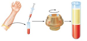
Plasma: 55% of blood; contains water, proteins, nutrients, hormones, and waste products.
Buffy Coat: <1% of blood; contains leukocytes (white blood cells) and platelets.
Erythrocytes: 45% of blood; red blood cells responsible for gas transport.
Physical Characteristics of Blood
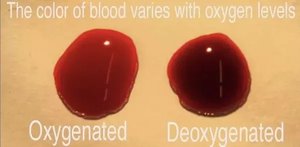
Blood is slightly alkaline (pH 7.35–7.45), more viscous than water, and its color varies with oxygenation.
Volume: 4–5 L in women, 5–6 L in men (about 8% of body weight).
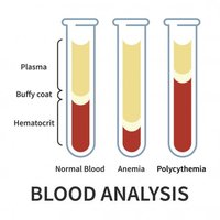
Hematocrit: Percentage of blood volume occupied by erythrocytes; normal values differ by gender.
Plasma
Composition and Function
Plasma is the clear, straw-colored liquid portion of blood, serving as the medium for transporting nutrients, hormones, and waste products.
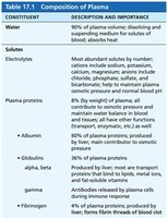
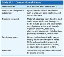
Constituent | Description and Importance |
|---|---|
Water | 90% of plasma volume; dissolving and suspending medium for solutes of blood; absorbs heat |
Electrolytes | Most abundant solutes; help maintain plasma osmotic pressure and normal blood pH |
Plasma proteins | 8% of plasma; includes albumin (osmotic pressure), globulins (transport, antibodies), and fibrinogen (clotting) |
Nonprotein nitrogenous substances | By-products of metabolism (urea, uric acid, creatinine) |
Nutrients | Glucose, amino acids, fatty acids, vitamins, etc. |
Respiratory gases | Oxygen and carbon dioxide |
Hormones | Steroid and thyroid hormones carried by plasma proteins |
Transport of Lipids in Plasma
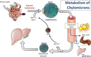
Lipids are insoluble in water and are transported in plasma bound to soluble plasma proteins, forming lipoproteins.
Formed Elements
Overview
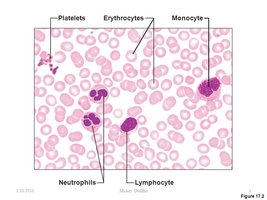
Formed elements include erythrocytes, leukocytes, and platelets. Most are short-lived and originate from stem cells in the bone marrow.
Erythrocytes (Red Blood Cells, RBCs)
Erythrocytes are specialized for oxygen and carbon dioxide transport. They are biconcave, anucleate cells packed with hemoglobin.
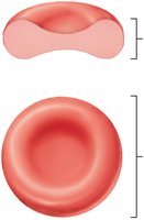
Shape: Biconcave disc increases surface area for gas exchange and flexibility for capillary passage.
Hemoglobin: Each RBC contains ~250 million hemoglobin molecules, each capable of binding four O2 molecules.
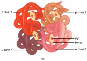
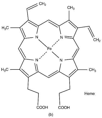
Oxyhemoglobin: Hemoglobin bound to oxygen (bright red).
Deoxyhemoglobin: Hemoglobin after oxygen release (dark red).
Carbaminohemoglobin: Hemoglobin bound to CO2.
Regulation of Erythropoiesis
Red blood cell production is regulated by erythropoietin (EPO), primarily produced by the kidneys in response to hypoxia (low oxygen levels).
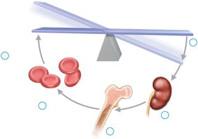
Essential nutrients: Iron, copper, zinc, folate, and vitamin B12 are required for erythropoiesis.
Disorders: Polycythemia (excess RBCs), anemia (deficiency in RBCs or hemoglobin).
RBC Life Cycle and Breakdown
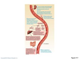
RBCs have a lifespan of about 120 days. Old or damaged RBCs are phagocytized in the liver, spleen, and bone marrow. Components are recycled or excreted.
Globin: Broken down into amino acids.
Iron: Stored as ferritin or hemosiderin, transported by transferrin.
Heme: Converted to biliverdin, then bilirubin, and excreted in bile.
RBC Disorders
Anemia: Reduced oxygen-carrying capacity due to blood loss, decreased production, or increased destruction of RBCs.
Polycythemia: Excess RBCs increase blood viscosity.
Hemolytic, sickle cell, and thalassemia: Genetic or acquired disorders affecting RBC structure or function.
Leukocytes (White Blood Cells, WBCs)
Types and Functions
Leukocytes are the only complete cells among the formed elements and are crucial for immune defense. They are classified as granulocytes or agranulocytes based on the presence of cytoplasmic granules.
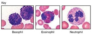
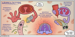
Granulocytes: Neutrophils, eosinophils, basophils
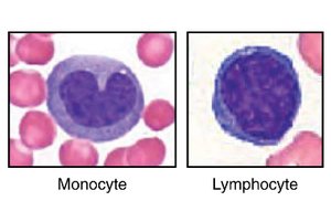
Agranulocytes: Lymphocytes, monocytes
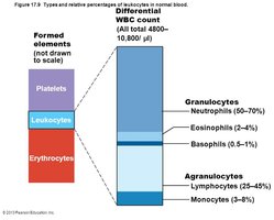
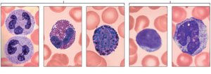
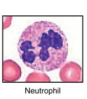
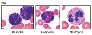
Neutrophils: Phagocytize bacteria; most abundant WBC.
Eosinophils: Combat parasitic infections and modulate allergic responses.
Basophils: Release histamine and heparin; involved in inflammation and allergic reactions.
Lymphocytes: B cells (antibody production), T cells (cell-mediated immunity), and natural killer cells.
Monocytes: Differentiate into macrophages for phagocytosis of pathogens and debris.
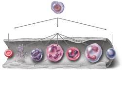
Leukopoiesis and Diapedesis
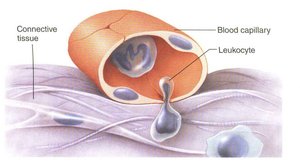
Leukocytes originate from hematopoietic stem cells in the bone marrow. They can exit the bloodstream via diapedesis to reach sites of infection or injury.
Platelets (Thrombocytes)
Structure and Function
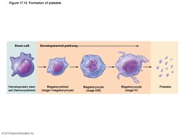
Platelets are cell fragments derived from megakaryocytes. They play a critical role in hemostasis by forming plugs at sites of vessel injury and releasing factors that promote clotting and tissue repair.
Normal count: 150,000–450,000/µL
Lifespan: ~10 days
Hemostasis
Steps of Hemostasis
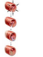
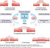
Hemostasis is the process of stopping blood loss from damaged vessels. It involves a series of tightly regulated steps:
Vascular Spasm: Vasoconstriction reduces blood flow.
Platelet Plug Formation: Platelets adhere to exposed collagen and aggregate.
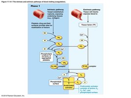
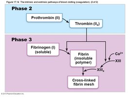
Coagulation: Cascade of clotting factors leads to fibrin mesh formation.
Clot Retraction and Fibrinolysis: Clot contracts and is eventually dissolved.
Clotting Disorders
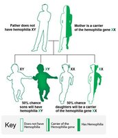
Hemophilia: Genetic deficiency of clotting factors (e.g., factor VIII in Hemophilia A).
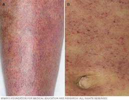
Thrombocytopenia: Low platelet count, leading to excessive bleeding and petechiae.
Thrombosis/Embolism: Excessive clot formation or dislodged clots causing vessel blockage.
Blood Groups and Transfusion
ABO Blood Group System
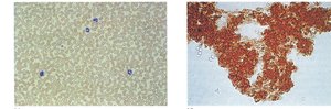
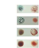
The ABO system classifies blood based on the presence of A and/or B antigens on erythrocytes. Mixing incompatible blood types causes agglutination (clumping) and potentially dangerous transfusion reactions.
Rh Blood Group System
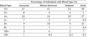
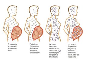
The Rh system is based on the presence (Rh+) or absence (Rh−) of the D antigen. Rh incompatibility can cause hemolytic disease of the newborn if an Rh− mother carries an Rh+ fetus.
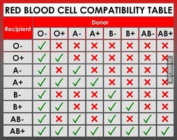
Summary Table: ABO and Rh Blood Types
Blood Type | Antigens on RBC | Antibodies in Plasma | Can Receive From | Can Donate To |
|---|---|---|---|---|
A | A | Anti-B | A, O | A, AB |
B | B | Anti-A | B, O | B, AB |
AB | A, B | None | A, B, AB, O | AB |
O | None | Anti-A, Anti-B | O | A, B, AB, O |
Additional info: Rh+ can receive Rh− blood once, but subsequent exposures may cause reactions due to antibody formation.
