 Back
BackBlood: Structure, Function, and Disorders – ANP College Study Guide
Study Guide - Smart Notes
 Tailored notes based on your materials, expanded with key definitions, examples, and context.
Blood – Physical Characteristics
Overview of Blood Properties
Blood is a specialized connective tissue with unique physical and chemical properties that are essential for its functions in the human body.
Volume: Adult males contain 5-6L; adult females contain 4-5L.
Temperature: Approximately 100.4°F, slightly higher than normal body temperature.
Viscosity: Blood is five times as viscous as water, making it more resistant to flow due to the presence of cells and plasma proteins.
pH: Ranges from 7.35 to 7.45, making it slightly alkaline.
Color: Varies from scarlet (oxygenated) to deep red (deoxygenated).
Components of Blood
Whole Blood Composition
Blood consists of plasma and formed elements, each with distinct roles.
Plasma: 46-63% of blood volume; contains water (92%), plasma proteins (7%), and other solutes (1%).
Formed Elements: 37-54% of blood volume; includes red blood cells (RBCs, 99.9%), white blood cells (WBCs), and platelets (0.1%).
Functions of Blood
Transportation
Blood is the primary medium for transporting substances throughout the body.
Gases: Oxygen and carbon dioxide are carried by hemoglobin and dissolved in plasma.
Nutrients: Absorbed from the GI tract and distributed to tissues.
Hormones: Transported from endocrine glands to target organs.
Metabolic Wastes: Carried to kidneys for excretion.
Regulation
Blood helps maintain homeostasis by regulating temperature, pH, and electrolyte balance.
Heat Distribution: Absorbs heat from muscles and distributes it to other tissues.
Acid-Base Balance: Neutralizes acids generated by active tissues.
Electrolyte Reservoir: Maintains levels of Na+, Ca2+, and other ions.
Volume Regulation: Adjusts in response to body water levels.
Protection
Blood provides defense against pathogens and restricts fluid loss.
White Blood Cells: Migrate to tissues to fight infections and remove debris.
Antibodies: Attack invading organisms and foreign compounds.
Clotting: Enzymes form clots to restrict fluid loss.
Red Blood Cells (Erythrocytes)
Structure and Function
Red blood cells are the most abundant formed elements and are specialized for oxygen transport.
Abundance: 4.5-6.3 million/µL in males; 4.2-5.5 million/µL in females.
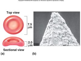
Shape: Biconcave disc (diameter ≈ 8µm, thickness ≈ 2.5µm) increases surface area for gas exchange and flexibility.
Organelles: Lack nucleus and most organelles; are membranous bags of hemoglobin.
Hemoglobin: Binds and transports O2 and CO2.
Hemoglobin Structure
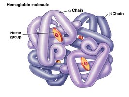
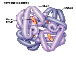
Hemoglobin is a large protein responsible for oxygen transport.
Composition: Four polypeptide chains (2 α, 2 β), each with a heme group containing iron.
Oxygen Binding: Each iron ion binds one O2 molecule; thus, each hemoglobin can carry four O2 molecules.
Red Blood Cell Formation (Erythropoiesis)
Process and Regulation
Erythropoiesis is the process of RBC formation, occurring in the red bone marrow.
Stem Cells: All blood cells develop from hemocytoblasts.
Pathways: Hemocytoblasts differentiate into myeloid and lymphoid stem cells, which give rise to RBCs, WBCs, and platelets.
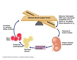
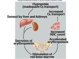
Regulation: Erythropoietin (EPO), produced by kidneys, stimulates RBC production in response to hypoxia.
Lifecycle and Fate of RBCs
Destruction and Recycling
RBCs have a lifespan of about 120 days and are subjected to mechanical stress.
Senescence: Old or damaged RBCs are engulfed by macrophages.
Hemolysis: RBCs rupture, and hemoglobin is broken down.
Fate of Components: Globin proteins are metabolized; heme is converted to bilirubin, which is transported to the liver and excreted in bile.
Bilirubin: Excess bilirubin causes jaundice; its breakdown products color urine and feces.
Complete Blood Count (CBC) and Hematocrit
Clinical Importance
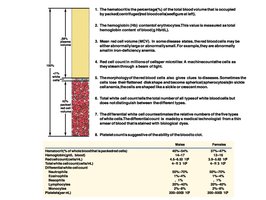
CBC is a routine test to determine the number and proportion of blood cells.
Hematocrit: Percentage of blood occupied by RBCs; average is 46% in males, 42% in females.
Clinical Significance: Low hematocrit indicates anemia; high values indicate polycythemia.
Blood Disorders
Polycythemia
Polycythemia is an elevated hematocrit with normal blood volume, often due to increased erythropoietin or bone marrow cancer.
Effects: Increases blood viscosity, reducing flow.
Treatment: Blood dilution with isotonic saline.
Application: Endurance athletes may induce polycythemia to enhance performance.
Anemia
Anemia is a condition of abnormally low oxygen-carrying capacity in the blood.
Symptoms: Shortness of breath, fatigue, chills.
Types: Hemorrhagic (blood loss), hemolytic (RBC rupture), aplastic (bone marrow destruction), iron-deficiency, pernicious (B12 deficiency), thalassemia, sickle cell.
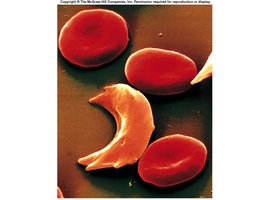
Sickle Cell Anemia
Sickle cell anemia is caused by a mutation in the β chain of hemoglobin, resulting in sickle-shaped RBCs that block blood vessels.
Genetic Basis: Single mutation in the globin gene.
Clinical Effects: Blockage of small vessels, pain, and organ damage.
Erythroblastosis Fetalis
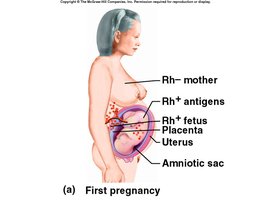
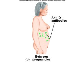
A type of hemolytic anemia affecting newborns when an Rh- mother produces antibodies against an Rh+ fetus.
Mechanism: Maternal antibodies attack fetal RBCs, causing anemia and hypoxia.
Prevention: Blood transfusions and Rh immunoglobulin therapy.
White Blood Cells (Leukocytes)
Functions and Types
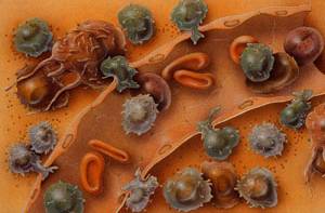
White blood cells defend the body against pathogens and remove toxins, wastes, and abnormal cells.
Abundance: 6000-9000/µL of blood.
Location: Most are in connective tissues and lymphatic organs.
Classification: Granulocytes (neutrophils, eosinophils, basophils) and agranulocytes (lymphocytes, monocytes).
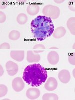
Granulocytes
Neutrophils: 50-70% of WBCs; first responders to infection; attack bacteria.
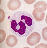
Eosinophils: 2-4% of WBCs; defend against parasites and participate in allergic reactions.
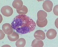
Basophils: <1% of WBCs; release histamine and heparin at injury sites.
Agranulocytes
Monocytes: 2-8% of WBCs; become macrophages in tissues and are aggressive phagocytes.
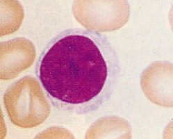
Lymphocytes: 20-30% of WBCs; include T cells (cellular immunity), B cells (antibody production), and NK cells (immune surveillance).
Platelets and Hemostasis
Platelet Function
Platelets are cell fragments involved in blood clotting and tissue repair.
Production: Released from megakaryocytes in bone marrow.
Lifespan: 9-12 days; removed by splenic phagocytes.
Abundance: 350,000/µL of blood.
Hemostasis (Blood Clotting)
Hemostasis prevents blood loss and establishes a framework for tissue repair.
Vascular Phase: Endothelial cells contract, release chemicals, and become sticky; vascular spasm reduces blood loss.
Platelet Phase: Platelets adhere to exposed surfaces and become activated, forming a platelet plug.
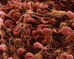
Coagulation Phase: Fibrinogen is converted to fibrin, trapping RBCs and platelets to form a clot.
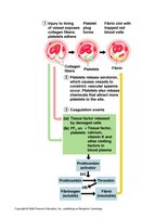
Clot Retraction and Manipulation
Clot retraction stabilizes the injury site and facilitates tissue repair. Anticoagulant drugs manipulate hemostasis to prevent unwanted clotting.
Clot Retraction: Platelets contract, pulling vessel edges together.
Anticoagulants: Heparin, Coumadin, Streptokinase, and Aspirin inhibit various steps in clot formation.
Thrombi, Emboli, and Hemophilia
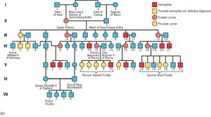
Thrombi are clots that form in intact vessels; emboli are clots that travel in the bloodstream. Hemophilia is a hereditary bleeding disorder due to deficiency of clotting factors.
Thrombus: Can obstruct blood flow and cause tissue damage.
Embolus: Can cause blockages in critical vessels, leading to stroke or pulmonary embolism.
Hemophilia: Most commonly due to lack of Factor VIII (Hemophilia A); X-linked inheritance.
Blood Type Compatibility
Blood Types and Transfusion
Blood type compatibility is crucial for safe transfusions.
Type O negative: Universal donor.
Type AB positive: Universal recipient.
Rh factor: Important in pregnancy and transfusion reactions.
Summary Table: Blood Cell Types and Functions
Cell Type | Abundance | Function |
|---|---|---|
Red Blood Cells | 99.9% of formed elements | Oxygen and carbon dioxide transport |
Neutrophils | 50-70% of WBCs | Bacterial defense |
Lymphocytes | 20-30% of WBCs | Immune response |
Monocytes | 2-8% of WBCs | Phagocytosis |
Eosinophils | 2-4% of WBCs | Parasite defense, allergy |
Basophils | <1% of WBCs | Histamine and heparin release |
Platelets | 350,000/µL | Clotting |
