 Back
Blood: Structure, Function, and Disorders – ANP College Study Guide
Study Guide - Smart Notes
 Tailored notes based on your materials, expanded with key definitions, examples, and context.
Blood: Structure and Function
Functions of Blood
Blood is a vital connective tissue responsible for transporting substances, regulating physiological parameters, and defending the body against disease.
Transport: Delivers oxygen (O2) and nutrients to tissues, removes carbon dioxide (CO2) and wastes, and transports hormones.
Regulation: Maintains temperature, pH, and extracellular fluid levels.
Protection: Clotting proteins close wounds; white blood cells defend against infection.
Characteristics of Blood
Blood exhibits specific physical and chemical properties that are essential for its function.
pH: 7.35–7.45
Temperature: 100.4º F
Volume: Males: 5–6 L; Females: 4–5 L
Color: Scarlet (high O2) to dark red (low O2)
Components of Blood
Blood consists of plasma and formed elements, each with distinct roles.
Plasma: Fluid matrix, ~90% water, contains proteins (albumin, globulins, fibrinogen), nutrients, electrolytes, gases, hormones, and wastes.
Formed Elements: Red blood cells (RBCs), white blood cells (WBCs), and platelets.
Hematocrit Procedure
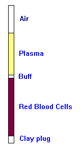
The hematocrit is a measure of the proportion of RBCs in blood, determined by centrifuging a blood sample.
Plasma: ~55% of blood volume
Buffy coat: Contains WBCs and platelets (<1% of blood volume)
RBCs: Hematocrit (Males: 47% ± 5%; Females: 42% ± 5%)
Plasma and Plasma Proteins
Composition of Plasma
Plasma is the liquid component of blood, serving as a transport medium for various substances.
Water: 90% of plasma
Proteins: Albumin (60%), globulins (alpha, beta, gamma), fibrinogen
Other Components: Nitrogenous wastes, nutrients, electrolytes, respiratory gases, hormones
Albumin
Albumin is the most abundant plasma protein, crucial for maintaining osmotic pressure and transporting substances.
Functions: Carrier protein, buffer, maintains water balance in blood
Red Blood Cells (Erythrocytes)
Structure and Function
Red blood cells are specialized for oxygen transport and exhibit unique structural features.
Shape: Biconcave discs, increasing surface area and facilitating gas exchange
Anucleate: Lack a nucleus
Hemoglobin: Packed with hemoglobin for O2 transport
No mitochondria: Do not consume the O2 they carry
Hemoglobin
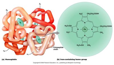
Hemoglobin is the protein responsible for binding and transporting oxygen and carbon dioxide.
Structure: 2 alpha and 2 beta chains, each with an iron (Fe) molecule
Oxygen Binding: Each hemoglobin molecule can carry 4 O2 molecules
RBC Content: Each RBC contains ~250 million hemoglobin molecules
RBC Vocabulary
Oxyhemoglobin: Oxygenated hemoglobin
Deoxyhemoglobin: Reduced hemoglobin (no O2)
Carbaminohemoglobin: CO2 bound to hemoglobin
RBC Counts
Women: 4.2–5.4 million cells/µL
Men: 4.7–6.1 million cells/µL
Viscosity: RBC count affects blood viscosity
Formation and Regulation of Blood Cells
Hematopoiesis
Hematopoiesis is the process of blood cell formation, occurring in red bone marrow.
Location: Axial skeleton, girdles, proximal epiphyses of humerus and femur in adults
Stem Cell: All blood elements derive from the hematopoietic stem cell (hemocytoblast)
Production Rate: ~100 billion new RBCs/day
Erythropoiesis (RBC Formation)
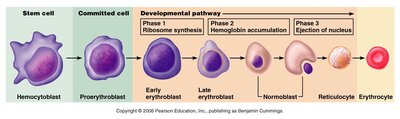
Erythropoiesis is the specific process of red blood cell formation, taking about 15 days.
Stages: Hemocytoblast → Proerythroblast → Early/Late erythroblast → Normoblast → Reticulocyte → Erythrocyte
Reticulocyte: Immature RBC that enters circulation and matures in 1–2 days
Regulation of Erythropoiesis
Regulation is primarily controlled by the hormone erythropoietin, released by the kidneys in response to low O2.
Triggers: Low RBC count, low hemoglobin, low O2 availability
Negative Feedback: High O2 turns off erythropoietin production
Diet and Erythropoiesis
Nutrients: Fats, sugars, proteins, vitamin B-12, folic acid, iron
DNA Synthesis: B-12 and folic acid are essential for rapidly dividing cells
Aging and Destruction of RBCs
RBCs have a limited lifespan and are destroyed by macrophages, with their components recycled or excreted.
Lifespan: 100–120 days
Destruction: Occurs mainly in the spleen; heme is broken down to bilirubin, globin to amino acids
Bilirubin: Released into blood, picked up by liver, secreted as bile, converted to urobilinogen and stercobilin (brown pigment)
RBC Disorders
Anemia
Anemia is a condition of low O2 carrying capacity, with various causes and symptoms.
Symptoms: Fatigue, pallor, shortness of breath, chills
Causes: Blood loss, RBC destruction, bone marrow failure, renal disease, iron/B12 deficiency, abnormal hemoglobin
Sickle Cell Anemia
Sickle cell anemia is an inherited disorder caused by a single amino acid substitution in the beta chain of hemoglobin.
Mechanism: Low O2 causes hemoglobin to change shape, resulting in sickled RBCs
Complications: Clotting, pain, stroke, infection
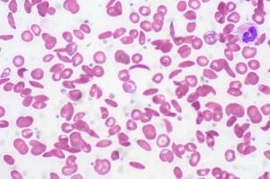
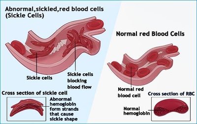
Population: Common in individuals of African descent; carriers are protected from malaria
Treatment: Transfusions, NO gas, drugs to activate fetal hemoglobin
Polycythemia
Polycythemia is a condition of excessive RBCs, increasing blood viscosity.
Causes: Bone cancer, high altitude, blood doping, synthetic EPO
White Blood Cells (Leukocytes)
Structure and Function
White blood cells are crucial for immune defense and exhibit unique behaviors.
Count: 4,800–10,800 WBCs/µL (<1% blood volume)
Diapedesis: Ability to leave the bloodstream
Amoeboid Motion: Movement outside the bloodstream
Chemotaxis: Attracted to sites by chemicals
Types of WBCs
Granulocytes: Neutrophils, eosinophils, basophils
Agranulocytes: Lymphocytes, monocytes
Granulocytes
Granulocytes have lobed nuclei and granules that stain with Wright’s stain; all are phagocytic to some degree.
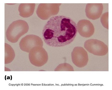
Neutrophils
Percentage: 50–70% of WBCs
Appearance: Small lilac granules, nucleus with 3–6 lobes
Function: Phagocytize bacteria; counts increase in bacterial infection
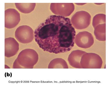
Eosinophils
Percentage: 2–4% of WBCs
Appearance: Red-staining granules, nucleus with 2 lobes
Function: Kill parasitic worms, reduce allergic reactions and asthma
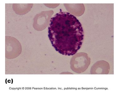
Basophils
Percentage: 0.5–1% of WBCs
Appearance: Large purple-black granules, S or U shaped nucleus
Function: Release histamine in inflammatory response
Agranulocytes
Agranulocytes lack membrane-bound granules.
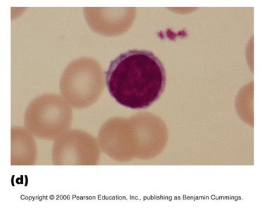
Lymphocytes
Percentage: ~25% of WBCs
Appearance: Large dark purple nucleus, pale blue cytoplasm
Function: Make antibodies, attack virus-infected and cancerous cells
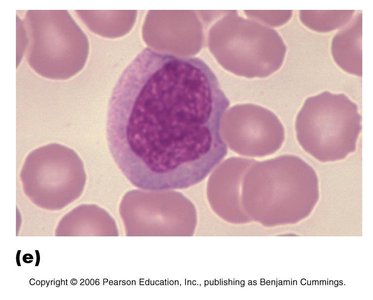
Monocytes
Percentage: 3–8% of WBCs
Appearance: Largest WBC, U or kidney-shaped nucleus, pale blue cytoplasm
Function: Phagocytosis; morph into macrophages in tissues
Mnemonic for WBC Order
"Never let monkeys eat bananas" helps remember the order of WBCs by number: Neutrophils, Lymphocytes, Monocytes, Eosinophils, Basophils.

Leukopoiesis and WBC Disorders
Leukopoiesis
Leukopoiesis is the production of WBCs, stimulated by interleukins and colony stimulating factors (CSF).
Lifespan: Granulocytes: hours–9 days; Agranulocytes: hours–decades
Pathways: Hemocytoblast → Myeloid stem cell (granulocytes, monocytes) or Lymphoid stem cell (lymphocytes)
WBC Disorders
Leukopenia: Too few WBCs; caused by drugs, diseases (HIV, lupus, influenza, malaria)
Leukemia: Excessive, nonfunctional WBCs; marrow fails to produce RBCs and platelets
Infectious Mononucleosis: Caused by Epstein-Barr virus; characterized by abnormal lymphocytes
Platelets and Hemostasis
Platelets
Platelets are cell fragments essential for blood clotting.
Count: 150,000–400,000/µL
Lifespan: ~10 days
Function: Contain granules with clotting chemicals
Formation of Platelets
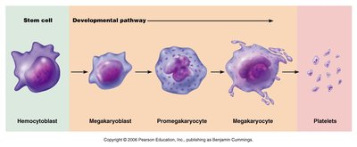
Platelet formation is stimulated by thrombopoietin and involves several developmental stages.
Pathway: Hemocytoblast → Megakaryoblast → Promegakaryocyte → Megakaryocyte → Platelets
Megakaryocyte: Releases platelets at the edge of sinusoid capillaries
Hemostasis
Hemostasis is the process of stopping bleeding, involving three main steps.
Vascular Spasms: Constriction of blood vessel to restrict blood loss
Platelet Plug Formation: Platelets adhere to exposed collagen, become sticky, and release chemicals
Coagulation: Blood transforms from liquid to gel; involves formation of prothrombin activator, conversion of prothrombin to thrombin, and thrombin converting fibrinogen to fibrin
Fibrin: Glues platelets together, turns plasma to gel, traps formed elements
Clotting: Complete in 3–6 min; stable in 30–60 min; fibrinolysis removes clot after healing
Healthy Vessels: Produce anti-clotting chemicals (heparin); blood flow dilutes clotting chemicals
Disorders of Hemostasis
Thromboembolic Disorders
Thrombus: Clot in unbroken vessel (e.g., coronary thrombosis)
Embolus: Clot that breaks away and circulates
Embolism: Clot stuck in small vessel (e.g., pulmonary, cerebral embolism)
Treatment: Drugs to dissolve/prevent clots (aspirin, heparin, warfarin)
Bleeding Disorders
Thrombocytopenia: Too few platelets; causes widespread hemorrhage
Impaired Liver Function: Liver makes clotting chemicals; vitamin K deficiency or liver failure
Hemophilia: Inherited disorder; treated with transfusions and clotting factor injections
Blood Types and Transfusions
Blood Types
Blood types are determined by the presence or absence of antigens (agglutinogens) on RBCs.
ABO System: Type A, B, AB, O
Antibodies: Develop against antigens not present on own RBCs
Blood Type | Antigens on RBC | Antibodies in Plasma | Blood Received |
|---|---|---|---|
A | A | Anti-B | A, O |
B | B | Anti-A | B, O |
AB (universal recipient) | A, B | None | A, B, AB, O |
O (universal donor) | None | Anti-A, Anti-B | O |
Rh Factor
Rh+: Antigen present
Rh-: Antigen absent; antibodies develop after exposure
Pregnancy: Rh- mother with Rh+ baby can develop antibodies, affecting subsequent pregnancies; prevented by RhoGAM
Blood Transfusions
Transfusion Reaction: Wrong blood type causes clots, RBC lysis, hemoglobin release, renal failure
Treatment: Fluids and diuretics to flush hemoglobin
Common Blood Tests
Types of Blood Tests
Total RBC Count/Hematocrit: Used to test for anemia
Differential WBC Count: Determines relative numbers of WBC types
Complete Blood Count (CBC): Counts all formed elements, hematocrit, hemoglobin content, RBC size
Complete Metabolic Profile (CMP): Provides blood chemistry, organ function, disease detection
Prothrombin Time and Platelet Counts: Assesses clotting function
Additional info: All explanations have been expanded for academic clarity and completeness, including inferred details about blood cell development, disorders, and clinical applications.
