Back
Blood: Structure, Function, and Laboratory Analysis
Study Guide - Smart Notes
 Tailored notes based on your materials, expanded with key definitions, examples, and context.
Blood: Structure and Function
Composition of Whole Blood
Blood is a fluid connective tissue composed of a liquid matrix (plasma) and suspended formed elements. Understanding its composition is essential for interpreting laboratory tests and physiological functions.
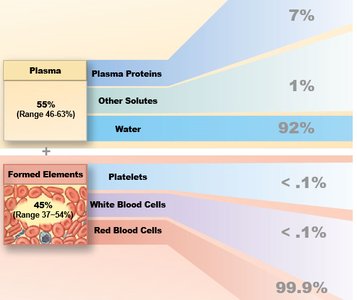
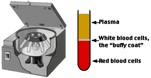
Plasma: The liquid portion of blood, making up about 55% of whole blood. Contains water, proteins (albumins, globulins, fibrinogen), nutrients, hormones, and waste products.
Formed Elements: Cellular components including erythrocytes (RBCs), leukocytes (WBCs), and platelets.
Major Constituents of Plasma
Plasma is primarily water (about 92%), with dissolved proteins and other solutes.
Plasma Proteins: Albumins (maintain osmotic pressure), globulins (immune function), fibrinogen (clotting).
Other Solutes: Electrolytes, nutrients, gases, hormones, and waste products.
Formed Elements of Blood
Types and Functions of Blood Cells
The formed elements include erythrocytes, platelets, and five types of leukocytes. Each has distinct structural features and functions.
Erythrocytes (RBCs): Biconcave, anucleate cells responsible for transporting oxygen and carbon dioxide.
Platelets: Small cytoplasmic fragments involved in blood clotting.
Leukocytes (WBCs): Immune cells classified as granulocytes (neutrophils, eosinophils, basophils) and agranulocytes (lymphocytes, monocytes).
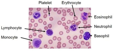
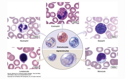
Classification and Identification of Leukocytes
Leukocytes are categorized based on their nuclear morphology and cytoplasmic granules. Their functions are vital for immunity and homeostasis.
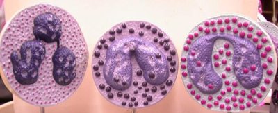
Neutrophils: Multi-lobed nucleus, most numerous (60-70%), phagocytose bacteria.
Lymphocytes: Large round nucleus, 20-30%, specific immunity (B and T cells).
Monocytes: Kidney-shaped nucleus, 3-8%, become macrophages in tissues.
Eosinophils: Red granules, 2-4%, combat parasitic infections and modulate allergic responses.
Basophils: Blue granules, 1%, release histamine, involved in inflammation and allergy.
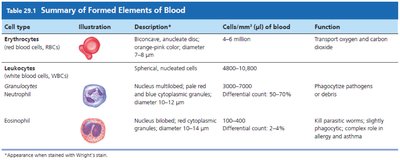
Summary Table: Formed Elements of Blood
The following tables summarize the key features, normal counts, and functions of blood cells.
Cell Type | Description | Normal Count (cells/mm3) | Function |
|---|---|---|---|
Erythrocytes | Biconcave, anucleate disc | 4-6 million | Transport O2 and CO2 |
Neutrophils | Multi-lobed nucleus, pale granules | 2500-7000 | Phagocytosis of pathogens |
Eosinophils | Bilobed nucleus, red granules | 100-400 | Kill parasites, modulate allergy |
Basophils | Lobed nucleus, blue granules | 20-50 | Release histamine, inflammation |
Lymphocytes | Round nucleus, scant cytoplasm | 1500-3000 | Specific immunity |
Monocytes | Kidney-shaped nucleus | 100-700 | Phagocytosis, become macrophages |
Platelets | Cytoplasmic fragments | 150,000-400,000 | Blood clotting |
Blood Tests and Laboratory Procedures
Hematocrit Test
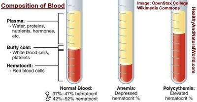
The hematocrit test measures the percentage of formed elements (mainly RBCs) in whole blood. It is a key indicator of blood health.
Formula:
Normal Values: Male: ~46%, Female: ~42%
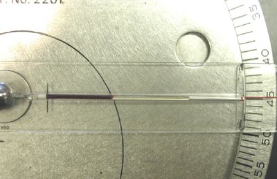
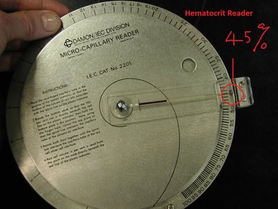
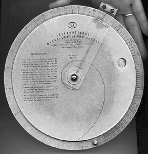
Procedure: Blood is placed in a capillary tube, sealed, and centrifuged. The proportion of RBCs is measured using a hematocrit reader.

Tallquist Hemoglobin Test
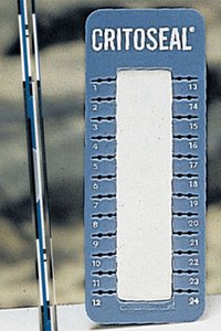
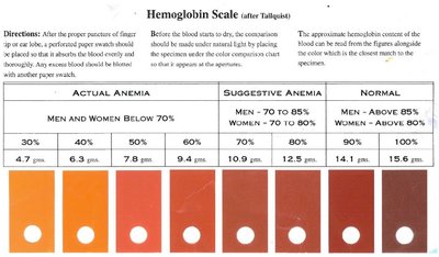
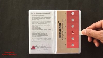
The Tallquist test estimates hemoglobin concentration by comparing the color of a blood stain to a standardized color scale.
Hemoglobin: Protein in RBCs that binds and transports O2 and CO2.
Normal Values: Adult male: 14–18 g/dL, Adult female: 12–16 g/dL
Procedure: Place a drop of blood on Tallquist paper, match color to scale, determine hemoglobin concentration.
Blood Type Tests: ABO and Rh Typing
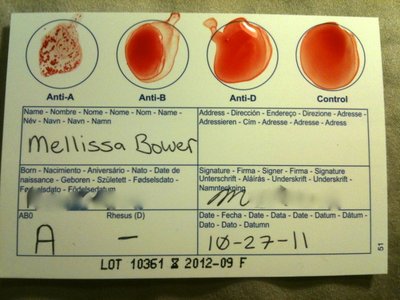
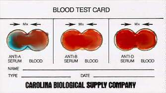
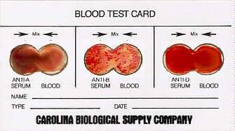
Blood typing identifies the presence of A, B, and Rh antigens on RBCs. Agglutination reactions with specific antisera determine blood type.
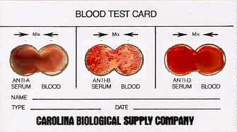
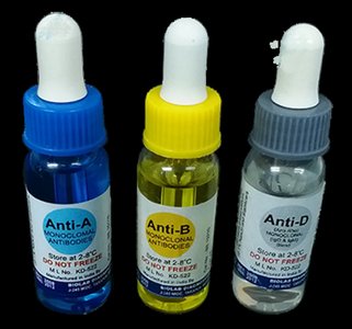
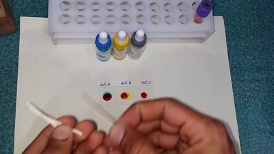
Testing Materials: Blood sample, Anti-A, Anti-B, and Anti-D (Rh) sera.
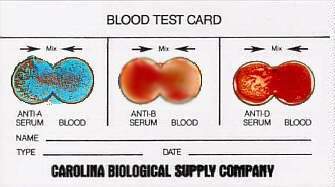
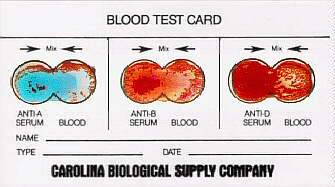
Procedure: Add each serum to separate spots, mix with blood, observe for agglutination (clumping).
Interpretation: Agglutination in a spot indicates presence of the corresponding antigen.
Example: Agglutination in A spot only = A- blood type.
Normal Values and Clinical Interpretation
Reference Ranges for Blood Cell Counts
RBC Count: 4-6 million cells/mm3
WBC Count: 4,000-10,800 cells/mm3
Platelet Count: 150,000-400,000 cells/mm3
WBC Differential: Neutrophils 60-70%, Lymphocytes 20-30%, Monocytes 3-8%, Eosinophils 2-4%, Basophils 1%
Clinical Significance
Low Hematocrit: May indicate anemia.
High Hematocrit: May indicate polycythemia.
Abnormal WBC Counts: May indicate infection, inflammation, or hematological disorders.
Summary
Blood is a complex tissue with critical roles in transport, immunity, and homeostasis. Laboratory tests such as hematocrit, hemoglobin, and blood typing are essential for clinical assessment and diagnosis.