 Back
BackBlood Vessel Physiology: Structure, Function, and Regulation
Study Guide - Smart Notes
 Tailored notes based on your materials, expanded with key definitions, examples, and context.
Blood Vessel Physiology
General Functions of Blood Vessels
Blood vessels are integral components of the cardiovascular system, responsible for transporting blood throughout the body. Their primary functions include:
Transport: Carrying blood to tissues for exchange of gases, nutrients, and wastes, then returning it to the heart.
Regulation: Controlling blood flow to tissues based on metabolic needs.
Pressure Control: Maintaining and regulating blood pressure within the circulatory system.
Chemical Secretion: Releasing substances that influence vascular tone and blood composition.
Arteries vs. Veins: Structure and Function
Arteries and veins are the two main types of blood vessels, each with distinct structural and functional characteristics:
Arteries: Carry blood away from the heart; have thick, muscular walls to withstand high pressure.
Veins: Return blood to the heart; have thinner walls and larger lumens, often equipped with valves to prevent backflow.
Key Structural Layers: Both arteries and veins have three layers:
Tunica intima (inner layer)
Tunica media (middle, muscular layer)
Tunica externa (outer layer)
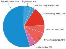
Blood Vessel Distribution
The cardiovascular system distributes blood unevenly among its components:
Systemic veins: Contain the majority of blood volume (~55%).
Arteries, capillaries, and heart: Hold smaller proportions.
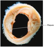
Atherosclerosis: Pathology and Impact
Atherosclerosis is a disease affecting large and medium-sized arteries, characterized by the formation of plaques within the tunica intima. These plaques consist of lipids, cholesterol, calcium salts, and cellular debris, and are often triggered by endothelial injury (e.g., high blood pressure, toxins, infections).
Impact: Plaques narrow the vessel lumen, reduce blood flow, and increase risk of cardiovascular events.
Inflammation: Vessel wall inflammation attracts phagocytes, which attempt to clear debris.
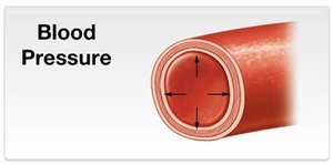
Blood Pressure: Definition and Determinants
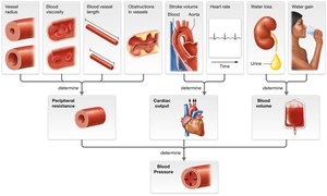
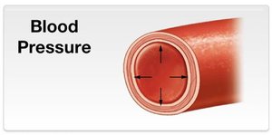
Blood pressure is the force exerted by blood on vessel walls, measured in millimeters of mercury (mm Hg). It varies across the vascular system, being highest in systemic arteries and lowest in veins.
Determinants:
Peripheral resistance (vessel radius, length, viscosity)
Cardiac output (heart rate, stroke volume)
Blood volume
Blood Pressure and Vessel Cross-Sectional Area
As arteries branch into smaller vessels, the total cross-sectional area increases, causing blood velocity to decrease. This slower flow in capillaries facilitates efficient exchange of gases and nutrients.
Example: If a large vessel has an area of 5 cm2 and branches into five smaller vessels each with 2 cm2, the total area becomes 10 cm2, doubling the original.
Blood Pressure Graph: Changes Across Vessels
Blood pressure declines as blood moves from arteries to veins. The graph shows pulsatile pressure in arteries, a sharp decline in arterioles, and continued decrease in capillaries and veins.
Venous Return: Skeletal and Respiratory Pumps
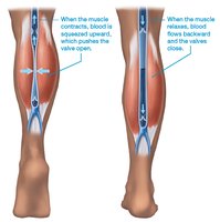
Veins rely on external mechanisms to return blood to the heart:
Skeletal muscle pump: Muscle contractions compress veins, pushing blood upward and opening valves.
Respiratory pump: Changes in thoracic and abdominal pressure during breathing create gradients that move blood toward the heart.
Neural and Hormonal Control of Blood Pressure
Blood pressure is regulated by short-term mechanisms involving the autonomic nervous system and hormones:
Sympathetic stimulation: Increases heart rate, contractility, and vasoconstriction, raising blood pressure.
Parasympathetic stimulation: Decreases heart rate and promotes vasodilation, lowering blood pressure.
Baroreceptor reflex: Sensors in the aorta and carotid arteries detect changes in pressure and trigger compensatory responses.
Chemoreceptor reflex: Responds to changes in blood oxygen, carbon dioxide, and pH, influencing vascular tone.
Capillary Structure and Function
Capillaries are the smallest blood vessels, specialized for exchange between blood and tissues. Their thin walls (single layer of endothelial cells) facilitate rapid diffusion of gases, nutrients, and wastes.
Structure: Composed of a lumen and a thin tunica intima.
Function: Efficient exchange due to minimal barrier.
Capillary Exchange Mechanisms
Exchange across capillary walls occurs via:
Diffusion: Movement of small molecules (e.g., O2, CO2) down concentration gradients.
Transcytosis: Transport of larger molecules via vesicles.
Bulk flow: Movement of water and solutes driven by pressure gradients.
Blood Flow Through Capillary Beds
Capillary beds are networks of capillaries supplied by arterioles and drained by venules. Blood flow is regulated by:
Precapillary sphincters: Rings of smooth muscle that control entry into capillaries.
Local and systemic factors: Including myogenic and metabolic mechanisms.
Local Regulation of Capillary Beds
Blood flow in capillary beds is adjusted by:
Myogenic mechanism: Changes in arteriolar resistance in response to pressure fluctuations.
Metabolic controls: Local chemical signals (O2, CO2, H+) cause vasodilation or vasoconstriction based on tissue activity.
Capillary Bed Pressures: Hydrostatic and Osmotic Forces
Movement of fluid in and out of capillaries is governed by:
Hydrostatic pressure: Pushes fluid out of capillaries into tissues.
Osmotic pressure: Draws fluid back into capillaries from tissues.
Net filtration pressure (NFP): Determines the direction and magnitude of fluid movement.
Key Equations
Blood Pressure:
Net Filtration Pressure (NFP):
Summary Table: Comparison of Arteries and Veins
Feature | Arteries | Veins |
|---|---|---|
Direction of Blood Flow | Away from heart | Toward heart |
Wall Thickness | Thick, muscular | Thin, less muscular |
Pressure | High | Low |
Valves | Absent | Present |
Lumen Size | Narrow | Wide |
Example: Clinical Measurement of Blood Pressure
Blood pressure is commonly measured using a sphygmomanometer, with readings given as systolic/diastolic values (e.g., 120/80 mm Hg).
Additional info:
These notes expand on brief lecture points to provide a comprehensive overview of blood vessel physiology, suitable for exam preparation in an anatomy and physiology college course.
