Back
BackBlood Vessel Structure, Function, and Circulatory Physiology
Study Guide - Smart Notes
 Tailored notes based on your materials, expanded with key definitions, examples, and context.
Blood Vessel Structure and Function
Overview of Blood Vessels
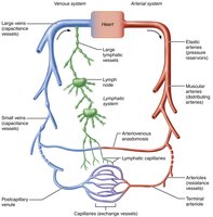
Blood vessels form a closed delivery system that begins and ends at the heart. They are dynamic structures that pulsate, constrict, relax, and even multiply, working with the lymphatic system to circulate fluids throughout the body. The three main types of blood vessels are arteries, capillaries, and veins, each with distinct roles in circulation.
Arteries: Carry blood away from the heart toward capillaries. Systemic arteries transport oxygenated blood, while pulmonary arteries carry oxygen-poor blood.
Capillaries: Serve as exchange vessels, allowing substances to move between blood and tissue cells.
Veins: Return blood from capillaries toward the heart. Systemic veins carry oxygen-poor blood, while pulmonary veins carry oxygenated blood.
Relationship of Blood Vessels and Lymphatic Vessels
The circulatory and lymphatic systems are closely linked. Lymphatic vessels collect excess interstitial fluid and return it to the bloodstream, helping maintain fluid balance and supporting immune function.
Blood Vessel Anatomy
General Structure of Blood Vessel Walls
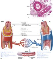
Except for capillaries, all blood vessels have three layers (tunics) surrounding a central lumen:
Tunica intima: Innermost layer, composed of endothelium (simple squamous epithelium) and a subendothelial layer in larger vessels. Provides a smooth, friction-reducing lining.
Tunica media: Middle layer, primarily smooth muscle and elastin. Responsible for vasoconstriction and vasodilation, regulating blood flow and pressure.
Tunica externa (adventitia): Outermost layer, mainly collagen fibers that protect, reinforce, and anchor the vessel. Contains nerves, lymphatics, and vasa vasorum (small vessels that nourish the outer wall).
Summary Table: Blood Vessel Anatomy
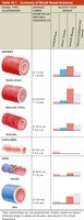
The following table summarizes the main structural and functional differences among the major types of blood vessels:
Vessel Type | Wall Thickness | Layers Present | Key Features |
|---|---|---|---|
Artery (Elastic) | Thick | All three | Large lumen, high elastin content, pressure reservoir |
Artery (Muscular) | Thick | All three | Thick tunica media, more smooth muscle, distributing vessel |
Arteriole | Thin | All three (smaller ones: mostly media and intima) | Major resistance vessels, control flow into capillaries |
Capillary | Very thin | Intima only | Exchange vessel, single endothelial layer |
Venule | Thin | Intima, thin media, thin externa | Very porous, allow WBCs/fluid movement |
Vein | Thinner than artery | All three | Large lumen, valves, capacitance vessel |
Types of Blood Vessels
Arteries: Pressure Reservoirs, Distributing Vessels, and Resistance Vessels
Elastic arteries: Largest arteries (e.g., aorta), act as pressure reservoirs, expand and recoil to maintain blood flow.
Muscular arteries: Distribute blood to specific organs, thick tunica media, more smooth muscle.
Arterioles: Smallest arteries, control flow into capillary beds via vasoconstriction and vasodilation, major determinants of resistance.
Capillaries: Exchange Vessels
Capillaries are the smallest blood vessels, consisting only of a thin tunica intima. Their primary function is the exchange of gases, nutrients, wastes, and hormones between blood and interstitial fluid. Most tissues have a rich capillary supply, except for poorly vascularized tendons, ligaments, cartilage, epithelia, cornea, and lens.
Types of Capillaries
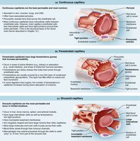
Continuous capillaries: Least permeable, most common, found in skin, muscles, lungs, and CNS. Endothelial cells joined by tight junctions with intercellular clefts for limited passage.
Fenestrated capillaries: Have pores (fenestrations) for increased permeability, found in kidneys, intestines, and endocrine glands.
Sinusoidal capillaries: Most permeable, found in liver, bone marrow, spleen, and adrenal medulla. Large clefts and fenestrations allow passage of large molecules and cells.
Capillary Beds
Capillary beds are networks of capillaries between arterioles and venules, providing sites for exchange with tissues. Blood flow through capillary beds is regulated by the diameter of arterioles and local chemical conditions.
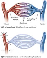
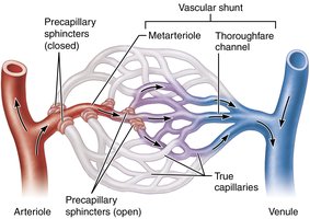
Specialized features in some beds include vascular shunts (direct connections between arterioles and venules) and precapillary sphincters (smooth muscle cuffs that regulate flow into true capillaries).
Veins: Blood Reservoirs
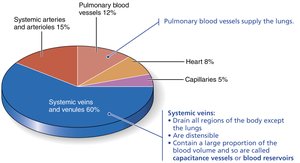
Veins return blood to the heart and act as capacitance vessels, containing up to 65% of the blood supply at any time. They have thinner walls and larger lumens than arteries, and blood pressure is lower. Adaptations such as large lumens and venous valves help ensure blood returns to the heart.
Physiology of Circulation
Blood Flow, Pressure, and Resistance
Blood flow is the volume of blood moving through a vessel, organ, or the entire circulation per unit time. Blood pressure is the force per unit area exerted on a vessel wall by the blood, and resistance is the opposition to flow, mainly due to friction within the vessel.
Blood viscosity: Increased viscosity increases resistance.
Blood vessel length: Longer vessels increase resistance.
Blood vessel diameter: Smaller diameter increases resistance exponentially (inversely proportional to the fourth power of the radius).
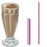
Relationship Between Flow, Pressure, and Resistance
The relationship is described by the equation:
Where F is blood flow, ΔP is the pressure gradient, and R is resistance. Blood flow is directly proportional to the pressure gradient and inversely proportional to resistance.
Blood Pressure Changes Throughout Circulation
Blood pressure is highest in the aorta and decreases through the systemic circulation, with the steepest drop in the arterioles. Capillary pressure is low to prevent rupture and allow exchange, while venous pressure is steady and low.
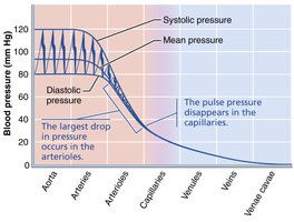
Arterial Blood Pressure
Systolic pressure: Peak pressure during ventricular contraction (about 120 mm Hg).
Diastolic pressure: Lowest pressure during ventricular relaxation (about 70–80 mm Hg).
Pulse pressure: Difference between systolic and diastolic pressure.
Mean arterial pressure (MAP): Average pressure propelling blood to tissues, calculated as:
Venous Return Mechanisms
Because venous pressure is low, three mechanisms assist venous return:
Muscular pump: Skeletal muscle contractions "milk" blood toward the heart.
Respiratory pump: Pressure changes during breathing move blood toward the heart.
Sympathetic venoconstriction: Reduces venous capacitance, pushing blood toward the heart.
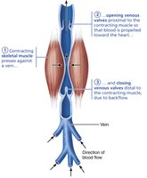
Blood Pressure Regulation
Major Factors Affecting Blood Pressure
Blood pressure is regulated by cardiac output (CO), total peripheral resistance (TPR), and blood volume. The relationship is:
Where:
CO (Cardiac Output) = Heart Rate (HR) × Stroke Volume (SV)
TPR is mainly determined by vessel diameter
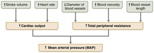
Short-Term Regulation: Neural and Hormonal Controls
Neural controls: Baroreceptor reflexes in carotid sinuses and aortic arch detect changes in pressure and adjust vessel diameter and heart rate accordingly.
Chemoreceptor reflexes: Respond to changes in blood CO2, pH, and O2 to adjust cardiac output and vessel tone.
Hormonal controls: Epinephrine, norepinephrine, angiotensin II, ADH, and ANP influence blood pressure by affecting vessel tone and blood volume.
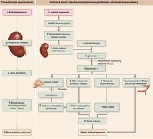
Long-Term Regulation: Renal Mechanisms
Kidneys regulate blood pressure by controlling blood volume through direct (urine formation) and indirect (renin-angiotensin-aldosterone system) mechanisms.
Intrinsic and Extrinsic Control of Blood Flow
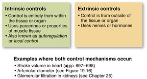
Intrinsic (Autoregulation) vs. Extrinsic Controls
Blood flow to tissues is regulated by:
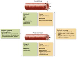
Intrinsic controls: Local mechanisms (metabolic and myogenic) that adjust blood flow to match tissue needs.
Extrinsic controls: Neural and hormonal mechanisms that maintain systemic blood pressure and redistribute blood during stress or exercise.
Blood Flow in Special Areas
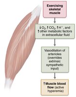
Skeletal muscle: Blood flow increases with activity due to metabolic autoregulation (active hyperemia).
Brain: Maintains constant flow; sensitive to pH and CO2 changes.
Skin: Regulates temperature and acts as a blood reservoir.
Lungs: Low pressure, unique autoregulation (low O2 causes vasoconstriction).
Heart: Blood flow increases during diastole and with increased activity.