 Back
BackBlood Vessels and Circulation: Structure and Function
Study Guide - Smart Notes
 Tailored notes based on your materials, expanded with key definitions, examples, and context.
Blood Vessels and Circulation
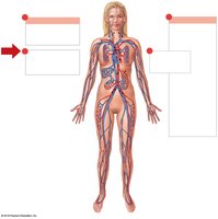
Overview of the Circulatory System
The circulatory system is responsible for transporting blood throughout the body, delivering oxygen and nutrients to tissues, and removing waste products. It consists of two main circuits: the pulmonary circuit (between the heart and lungs) and the systemic circuit (between the heart and the rest of the body).
Structure of Blood Vessels
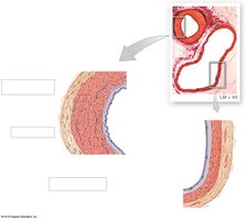
Layers of Blood Vessel Walls
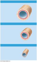
Blood vessels are composed of three main layers, or tunics, each with distinct structural and functional properties:
Tunica intima (interna): The innermost layer, consisting of endothelium and a basement membrane. In arteries, it includes an internal elastic membrane for elasticity.
Tunica media: The middle layer, primarily made of smooth muscle and elastic fibers. It is usually the thickest layer in arteries and is responsible for vasoconstriction and vasodilation.
Tunica externa (adventitia): The outermost layer, composed of connective tissue that provides structural support and elasticity.
Comparison of Arteries and Veins
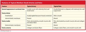
Arteries and veins differ in structure and function. The table below summarizes the main differences between typical medium-sized arteries and veins:
Feature | Typical Artery | Typical Vein |
|---|---|---|
General appearance in sectional view | Usually round, with relatively thick wall and small lumen | Usually flattened or collapsed, with relatively thin wall and large lumen |
Tunica intima | Endothelium usually rippled because of vessel constriction; internal elastic membrane present | Endothelium often smooth; internal elastic membrane absent |
Tunica media | Thick, dominated by smooth muscle cells and elastic fibers; external elastic membrane present | Thin, dominated by smooth muscle cells and collagen fibers; external elastic membrane absent |
Tunica externa | Collagen and elastic fibers | Collagen, elastic fibers, and smooth muscle cells |
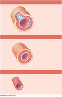
Types of Arteries and Veins
Blood vessels are classified based on their size and function:
Arteries: Carry blood away from the heart. Types include elastic arteries (large, conducting), muscular arteries (distribution), and arterioles (resistance vessels).
Veins: Return blood to the heart. Types include venules (smallest), medium-sized veins, and large veins.
Capillaries: Structure and Function
Types of Capillaries
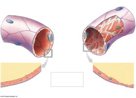
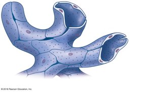
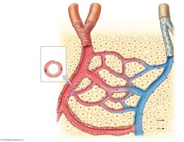
Capillaries are the smallest blood vessels and are the primary site of exchange between blood and tissues. There are three main types:
Continuous capillaries: Most common; endothelial cells form a continuous tube with small intercellular gaps. Found in muscle, connective tissue, brain, and lungs.
Fenestrated capillaries: Have pores (fenestrations) that allow rapid exchange of water and small solutes. Found in kidneys, endocrine glands, and intestines.
Sinusoids: Have large gaps and fenestrations, allowing passage of large molecules and cells. Found in liver, bone marrow, and spleen.

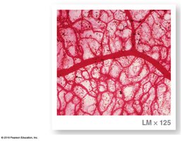
Capillary Beds and Blood Flow Regulation
Capillaries form networks called capillary beds, where blood flow is regulated by precapillary sphincters. These sphincters control the amount of blood entering the capillary bed, responding to local tissue needs and signals from the nervous system.
Venous System and Blood Distribution
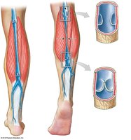
Venous Valves and Blood Return
Veins, especially in the limbs, contain valves that prevent backflow of blood. The contraction of surrounding skeletal muscles helps propel blood toward the heart, a mechanism known as the skeletal muscle pump.
Blood Reservoirs
At rest, most of the blood volume is found in systemic veins and venules, which act as blood reservoirs. These vessels can constrict (venoconstriction) to redistribute blood to other parts of the body during times of need, such as hemorrhage or increased activity.
Summary Table: Features of Arteries and Veins
Feature | Typical Artery | Typical Vein |
|---|---|---|
General appearance in sectional view | Usually round, with relatively thick wall and small lumen | Usually flattened or collapsed, with relatively thin wall and large lumen |
Tunica intima | Endothelium usually rippled because of vessel constriction; internal elastic membrane present | Endothelium often smooth; internal elastic membrane absent |
Tunica media | Thick, dominated by smooth muscle cells and elastic fibers; external elastic membrane present | Thin, dominated by smooth muscle cells and collagen fibers; external elastic membrane absent |
Tunica externa | Collagen and elastic fibers | Collagen, elastic fibers, and smooth muscle cells |
Key Terms and Concepts
Artery: Blood vessel that carries blood away from the heart.
Vein: Blood vessel that returns blood to the heart.
Capillary: Microscopic vessel where exchange of substances occurs between blood and tissues.
Tunica intima, media, externa: The three layers of blood vessel walls.
Precapillary sphincter: Ring of smooth muscle that regulates blood flow into capillary beds.
Venous valve: Structure in veins that prevents backflow of blood.
