 Back
BackBlood Vessels and Circulation: Structure, Function, and Regulation
Study Guide - Smart Notes
 Tailored notes based on your materials, expanded with key definitions, examples, and context.
Blood Vessels: Classification and Structure
Types of Blood Vessels
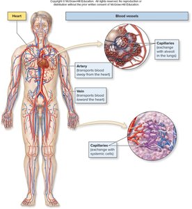
The cardiovascular system consists of three main types of blood vessels: arteries, veins, and capillaries. Each type plays a distinct role in the transport of blood throughout the body.
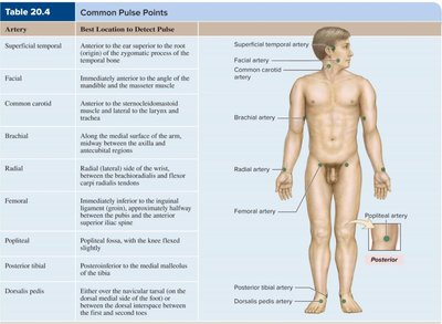
Arteries: Transport blood away from the heart. Typically carry oxygen-rich blood in systemic circulation.
Veins: Transport blood toward the heart. Typically carry oxygen-poor blood in systemic circulation.
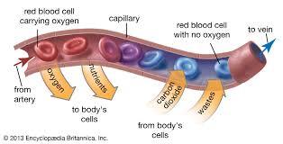
Capillaries: Facilitate exchange of gases, nutrients, and wastes between blood and tissues.
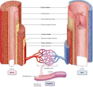
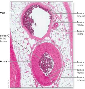
Structure of Blood Vessel Walls
Arteries and veins are composed of three layers, called tunics, surrounding a central lumen. Capillaries have a simpler structure.
Tunica Externa: Outer connective tissue layer; anchors vessel to surrounding tissues.
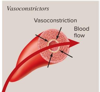
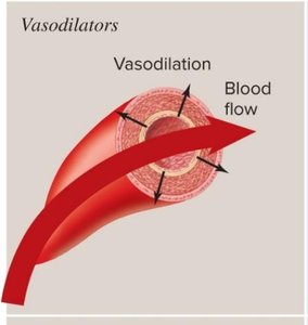
Tunica Media: Middle layer; contains smooth muscle and elastic fibers, responsible for vasoconstriction and vasodilation.
Tunica Intima: Inner layer; consists of endothelium (simple squamous epithelium) and provides a smooth surface for blood flow.
Capillaries: Composed only of endothelium and a basement membrane, allowing efficient exchange.
Companion Vessels
Arteries and veins that supply and drain the same body region are called companion vessels. They travel together, ensuring efficient circulation.
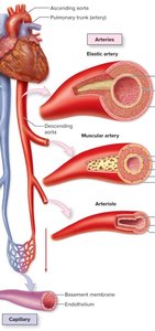
Classification of Arteries, Veins, and Capillaries
Arteries
Elastic Arteries: Largest arteries, high elastic content, near the heart (e.g., aorta).
Muscular Arteries: Medium-sized, distribute blood to specific regions (e.g., brachial artery).
Arterioles: Smallest arteries, regulate blood flow and pressure via smooth muscle contraction.
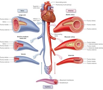
Veins
Large Veins: Largest veins, act as blood reservoirs (e.g., superior/inferior vena cava).
Small to Medium Veins: Companion to muscular arteries.
Venules: Smallest veins, drain capillaries.
Valves: Present in veins to prevent backflow due to low pressure.

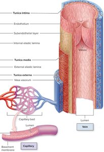
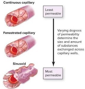
Capillaries
Continuous Capillaries: Least permeable, most common.
Fenestrated Capillaries: Slightly permeable, found in endocrine glands.
Sinusoid Capillaries: Most permeable, found in red bone marrow.
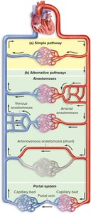
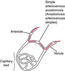
Pathways of Blood Vessels
Simple Pathway, Anastomosis, and Portal System
Simple Pathway: Blood flows from artery to capillary to vein.
Anastomosis: Joining of multiple arteries or veins; can provide alternate routes.
Portal System: Blood flows from one capillary bed to another via a portal vein (e.g., intestines to liver).
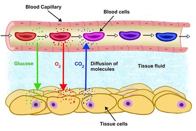
Capillary Exchange
Mechanisms of Exchange
Capillary exchange is the process by which substances move between blood and tissues. Three main mechanisms are involved:
Diffusion: Movement from high to low concentration (e.g., oxygen, glucose in; carbon dioxide out).
Vesicular Transport: Endocytosis and exocytosis move large molecules and proteins.
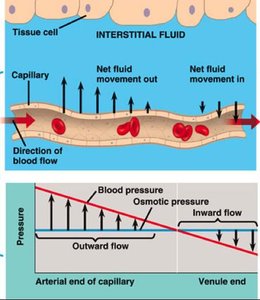
Bulk Flow: Blood pressure moves water and solutes into tissues; osmosis returns water to blood.
Forces Governing Bulk Flow
Hydrostatic Pressure: Pushes water and solutes out of capillaries at arterial end (filtration).
Osmotic Pressure: Pulls water into capillaries at venous end (reabsorption).
Degree of Vascularization and Angiogenesis
Vascularization
Highly Vascularized Tissues: Brain, skeletal muscle, heart, liver.
Poorly Vascularized Tissues: Tendons, ligaments, cartilage.
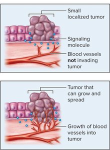
Angiogenesis and Regression
Angiogenesis: Formation of new blood vessels, occurs during tissue growth and in tumors.
Regression: Reduction in blood vessels, occurs during weight loss or decreased tissue demand.
Blood Pressure and Blood Flow
Blood Pressure
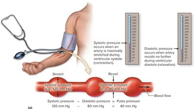
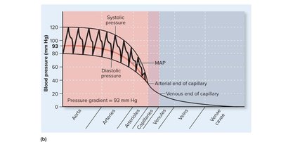
Blood pressure is the force exerted by blood against vessel walls, measured in mmHg. It is highest in the aorta and lowest in the vena cava.
Systolic Pressure: Maximum pressure during ventricular contraction.
Diastolic Pressure: Minimum pressure during ventricular relaxation.
Pulse Pressure: Difference between systolic and diastolic pressure; optimal value is 40 mmHg.
Blood Flow
Blood Flow: Amount of blood moving through the cardiovascular system per unit time.
Cardiac Output: Amount of blood pumped by the heart per minute.
Regulation of Blood Pressure and Flow
Factors Affecting Blood Pressure
Cardiac Output: Increased heart rate and stroke volume raise blood pressure.
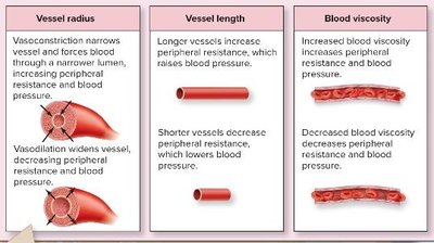
Peripheral Resistance: Resistance due to vessel radius, length, and blood viscosity.
Blood Volume: More blood increases pressure.
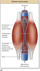
Venous Return
Skeletal Muscle Pump: Muscle contractions help push blood back to the heart.
Respiratory Pump: Breathing assists venous return.
Systemic Regulation
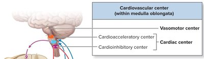
Cardiovascular Center: Located in the brainstem; includes vasomotor and cardiac centers.
Baroreceptors: Detect changes in blood pressure.
Chemoreceptors: Detect changes in blood chemistry (e.g., oxygen levels).
Autonomic Nervous System: Sympathetic and parasympathetic divisions regulate vessel tone and cardiac output.
Redistribution of Blood Flow During Exercise
Sympathetic Response
Epinephrine: Causes vasoconstriction in non-essential organs and vasodilation in skeletal muscles and heart.
Renin-Angiotensin System: Hormonal regulation increases blood pressure.
Antidiuretic Hormone (ADH): Promotes vasoconstriction to maintain blood pressure.
Pulmonary Circulation
Blood Pressure in Pulmonary Circuit
Pulmonary Blood Pressure: Lower than systemic circulation, reducing resistance and facilitating gas exchange.
Systemic Circulation and Vascular Diseases
Major Vessels and Regions
Head, Neck, and Arms: Supplied by branches of the aortic arch; drained by brachiocephalic veins.
Thoracic and Abdominal Organs: Supplied by descending aorta and its branches.
GI Tract: Supplied by celiac trunk and mesenteric arteries; drained by hepatic portal system.
Kidneys: Supplied by renal arteries and veins.
Lower Limbs: Supplied by common iliac arteries; drained by common iliac veins.
Vascular Diseases
Arteriosclerosis: Hardening and loss of elasticity in arteries; increases resistance and restricts blood flow.
Atherosclerosis: Build-up of plaque in arterial walls; primary cause of coronary artery disease.
Varicose Veins: Valves fail, causing blood to pool and veins to enlarge.
Cerebral Edema: Excess fluid in the brain due to high blood pressure.
Stroke: Reduced or blocked blood flow to the brain.
Peripheral Vascular Disease: Disorders affecting circulation outside the brain and heart.
Hypertension: Chronic high blood pressure; risk factors include diet, lifestyle, age, and genetics.
Treatments
Angioplasty: Surgical repair or unblocking of a blood vessel.
Medications: Blood thinners, vasodilators, and clot-busting drugs.
Lifestyle Changes: Diet and exercise to reduce risk factors.
Summary Table: Differences Between Arteries and Veins
Characteristic | Artery | Vein |
|---|---|---|
Lumen diameter | Narrow | Large |
Wall thickness | Thick | Thin |
Blood flow | Away from heart | Towards heart |
Blood pressure | Higher | Lower |
Elastic fibers | More | Less |
Blood oxygen | High (systemic) | Low (systemic) |
Valves | No | Yes |
Key Equations
Blood Pressure Gradient:
Pulse Pressure:
Cardiac Output:
Conclusion
The structure and function of blood vessels are essential for maintaining proper circulation and tissue health. Understanding the differences between arteries, veins, and capillaries, as well as the mechanisms regulating blood pressure and flow, is fundamental for studying the cardiovascular system and its associated diseases.
