 Back
BackBlood Vessels and Circulation: Structure, Function, and Regulation
Study Guide - Smart Notes
 Tailored notes based on your materials, expanded with key definitions, examples, and context.
Blood Vessels and Circulation
Overview of the Cardiovascular System
The cardiovascular system is composed of the heart (pump), blood vessels (conducting system), and blood (fluid medium). Blood circulates throughout the body, delivering oxygen and nutrients while removing waste products. Blood vessels are classified by their size and histological organization, and they play a crucial role in maintaining homeostasis.
Arteries: Carry blood away from the heart; the largest are the aorta and pulmonary trunk.
Arterioles: Smallest branches of arteries, with distinct functional and structural differences.
Capillaries: Smallest blood vessels; site of exchange between blood and interstitial fluid.
Venules: Collect blood from capillaries; small veins.
Veins: Return blood to the heart.
Structure of Blood Vessel Walls
Blood vessel walls are composed of three layers, each with distinct functions:
Tunica intima: Inner layer; includes the endothelial layer, connective tissue, and (in arteries) an internal elastic membrane.
Tunica media: Middle layer; contains smooth muscle cells in loose connective tissue. Thickest in arteries and contains an external elastic membrane.
Tunica externa: Outer layer; anchors the vessel to surrounding tissues.
Vasa vasorum are small blood vessels that supply the walls of larger arteries and veins.
Differences Between Arteries and Veins
Arteries have thicker walls and higher blood pressure than veins.
Arteries have a small, round lumen; veins have a large, flat lumen.
Arteries are more elastic and snap back to shape if distorted.
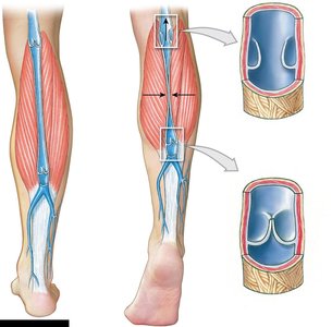
Veins have valves (formed by folds of the tunica intima) to prevent backflow of blood.
Types of Arteries and Capillaries
Elastic (conducting) arteries: Largest arteries; walls are resilient due to high density of elastic fibers in the tunica media. Elasticity evens out pulse force.
Muscular (distribution) arteries: Medium-sized; tunica media has many muscle cells.
Arterioles: Small; little or no tunica externa and a thin/incomplete tunica media. Diameter changes affect resistance to blood flow.
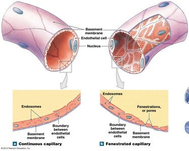
Capillaries are the smallest vessels with thin walls, permeating all active tissue. They lack tunica externa and tunica media. There are two major types:
Continuous capillaries: Complete endothelial lining; found in all tissues except epithelia and cartilage. Permit diffusion of water and small solutes.
Fenestrated capillaries: Have pores in the endothelial lining; permit rapid exchange of water and large solutes.
Veins and Venous Return
Veins collect blood from capillaries and return it to the heart. They are larger in diameter, have thinner walls, and lower blood pressure than arteries. Veins often contain valves to prevent backflow, especially in the limbs.
Pressure, Resistance, and Blood Flow
Blood Pressure and Resistance
Blood flow through capillaries is determined by pressure (P) and resistance (R). The heart generates pressure to overcome resistance. Key pressures include:
Blood pressure (BP): Arterial pressure, measured in mm Hg.
Capillary hydrostatic pressure (CHP): Pressure exerted by blood against capillary walls.
Venous pressure: Pressure within the venous system; typically low.
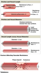
Total peripheral resistance is influenced by:
Vascular resistance: Primarily due to friction; depends on vessel length and diameter.
Blood viscosity: Resistance caused by interactions between molecules in blood.
Turbulence: Increases resistance; can be caused by plaques.
Arterial and Venous Pressure
Systolic pressure: Peak blood pressure during ventricular systole.
Diastolic pressure: Minimum pressure at the end of diastole.
Venous return is assisted by muscular compression and the respiratory pump.
Capillary Exchange
Capillary exchange is essential for tissue health and involves three processes:
Diffusion: Movement of ions and molecules from high to low concentration.
Filtration: Driven by hydrostatic pressure; water and small solutes are forced through the capillary wall.
Reabsorption: Result of osmotic pressure; brings water back into the capillary.
Continuous movement of water out of capillaries, through tissues, and back via reabsorption is critical for normal function.
Edema
Edema is the abnormal accumulation of interstitial fluid, caused by an imbalance between hydrostatic and osmotic forces at the capillary level. Causes include capillary damage, starvation (low plasma proteins), and high blood pressure.
Regulation of Blood Flow and Pressure
Cardiovascular Regulation
Blood flow is regulated to meet tissue needs without compromising vital organs. Mechanisms include:
Autoregulation: Immediate, localized adjustments.
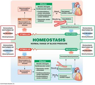
Neural mechanisms: Rapid responses coordinated by the cardiac and vasomotor centers of the medulla oblongata.
Endocrine mechanisms: Direct, long-term changes via hormones.
Hormonal Regulation
Antidiuretic hormone (ADH): Elevates BP, reduces water loss at kidneys.
Angiotensin II: Responds to low renal BP; increases cardiac output and stimulates other hormones.
Erythropoietin (EPO): Released in response to low BP and low O2 in blood.
Natriuretic peptides (ANP, BNP): Lower BP and blood volume in response to excessive stretching.
Cardiovascular Adaptation and Circuits
Adaptation to Stress
The cardiovascular system adapts to stress through coordinated responses during exercise, hemorrhage, and by prioritizing blood flow to the brain, heart, and lungs.
Exercise: Vasodilation in muscles, increased cardiac output, restricted flow to non-essential organs.
Hemorrhage: Vasoconstriction and hormonal responses to maintain BP and restore blood volume.
Special Circulations: Brain, heart, and lungs have unique regulatory mechanisms to ensure constant supply.
Pulmonary and Systemic Circuits
The cardiovascular system is divided into pulmonary (lungs) and systemic (rest of body) circuits. Peripheral artery and vein distribution is generally symmetrical, and tissues are often supplied by multiple vessels interconnected by anastomoses.
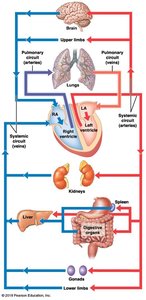
Pulmonary Circuit
Deoxygenated blood from the systemic circuit enters the right atrium and ventricle, then the pulmonary trunk, which branches into pulmonary arteries and capillaries around alveoli. Gas exchange occurs, and oxygenated blood returns to the left atrium via pulmonary veins.
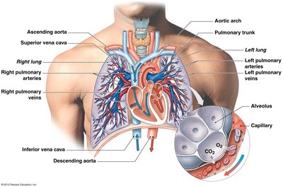
Systemic Circuit
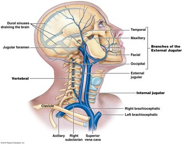
The systemic circuit supplies all body tissues except the lungs. Major arteries branch from the aorta, and veins return blood to the right atrium via the superior and inferior vena cava. Arteries are typically paired and named by region; veins show more individual variation and often have superficial and deep sets.
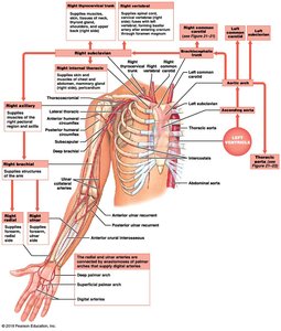
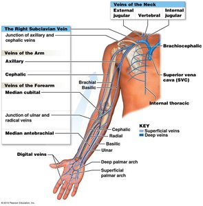
Major Arteries and Veins of the Trunk and Limbs
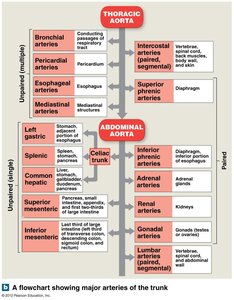
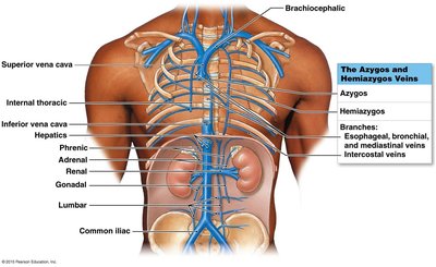
The aorta branches into the thoracic and abdominal aorta, supplying various organs and tissues. The common iliac arteries supply the legs, branching into internal and external iliac arteries. Major veins include the azygos and hemiazygos systems, which drain the thoracic and abdominal walls.
Fetal Circulation
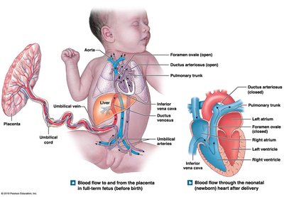
Fetal circulation differs from adult circulation due to non-functional lungs. Oxygen and nutrients are received via the placenta. The umbilical vein carries oxygenated blood to the fetus, and the foramen ovale and ductus arteriosus connect the right and left sides of the heart. After birth, these structures close as the lungs become functional.
Aging and the Cardiovascular System
With age, the cardiovascular system undergoes several changes:
Decreased hematocrit and pooling of blood in leg veins.
Reduction in cardiac output and changes in nodal/conducting cells.
Progressive atherosclerosis and less elastic arteries.
Calcium deposits on vessel walls and scar tissue in cardiac muscle.
