Back
BackBlood Vessels and Circulation: Structure, Function, and Regulation
Study Guide - Smart Notes
 Tailored notes based on your materials, expanded with key definitions, examples, and context.
Blood Vessels and Circulation
Classes of Blood Vessels
The cardiovascular system consists of several classes of blood vessels, each with distinct structural and functional characteristics.
Arteries: Carry blood away from the heart; branch repeatedly into smaller arteries.
Arterioles: Smallest branches of arteries; lead to capillary beds.
Capillaries: Smallest blood vessels; site of exchange between blood and interstitial fluid.
Venules: Smallest branches of veins; collect blood from capillaries.
Veins: Return blood to the heart; smaller veins unite to form larger veins.
Structure of Vessel Walls
Blood vessel walls are composed of three layers, each contributing to the vessel's function and integrity.
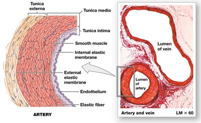
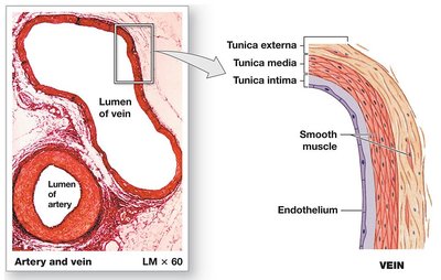
Tunica intima: Inner layer; lines the lumen and includes an endothelial lining surrounded by connective tissue and elastic fibers. In arteries, the internal elastic membrane is present.
Tunica media: Middle layer; contains concentric sheets of smooth muscle in a framework of loose connective tissue. The external elastic membrane separates it from the tunica externa.
Tunica externa (adventitia): Outer layer; contains connective tissue, collagen, and elastic fibers. In veins, it is thicker and contains smooth muscle. The vasa vasorum supplies cells of the tunica media and externa in large vessels.


Differences Between Arteries and Veins
Arteries and veins differ in structure and function due to the pressures they encounter and their roles in circulation.
Arteries: Thicker walls, more smooth muscle and elastic fibers, maintain circular shape, pleated endothelium when constricted.
Veins: Larger lumen, collapse when sectioned, have valves to prevent backflow (important for working against gravity).


Representative Views of Vessel Types
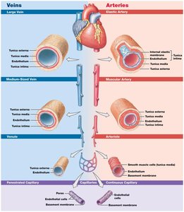
Diagrammatic views illustrate the structural differences between arteries, veins, and capillaries.
Arterial Structure and Function
Arteries can change diameter passively (elasticity) and actively (autonomic control).
Vasoconstriction: Contraction of arterial smooth muscle, reducing diameter.
Vasodilation: Relaxation of arterial smooth muscle, increasing diameter.
These changes affect afterload, peripheral blood pressure, capillary blood flow, and hemostasis.
Types of Arteries
Elastic arteries: Largest, closest to the heart; resilient walls with many elastic fibers; exhibit elastic rebound.
Muscular arteries: Medium-sized; thick tunica media with much smooth muscle; important for pressure points.
Arterioles: Smallest arteries; thin or incomplete tunica media; called resistance vessels due to their role in regulating resistance.
Clinical Conditions Affecting Arteries
Aneurysm: Bulge in a weakened arterial wall; risk of rupture and internal bleeding.
Arteriosclerosis: Hardening and thickening of arterial wall.
Atherosclerosis: Lipid deposits (plaque) inside artery; can lead to focal calcification (calcium salt deposition).
Capillaries: Structure and Types
Capillaries are the site of exchange between blood and interstitial fluid. Their thin walls consist only of tunica intima.
Continuous capillaries: Complete endothelial lining; found in most tissues except epithelia and cartilage; restricted permeability in CNS and thymus (e.g., blood-brain barrier).
Fenestrated capillaries: Pores in endothelial lining; permit exchange of water and larger solutes; found in choroid plexus, endocrine organs, kidneys, intestinal tract.
Sinusoids: Gaps between endothelial cells; permit exchange of water and large plasma proteins; found in liver, spleen, bone marrow, endocrine organs; monitored by phagocytic cells.
Capillary Beds and Blood Flow Regulation
Capillary beds are networks of interconnected capillaries. Blood flow is regulated by precapillary sphincters and thoroughfare channels.
Precapillary sphincters: Smooth muscle cells controlling blood flow through capillaries.
Thoroughfare channels: Direct passageways between arterioles and venules.
Anastomoses: Interconnections between arteries or between arterioles and venules, allowing alternative routes for blood flow.
Angiogenesis: Formation of new blood vessels, stimulated by vascular endothelial growth factor (VEGF).
Veins: Structure and Function
Veins collect blood from capillaries and return it to the heart. They have all three vessel wall layers and are classified by size.
Venules: Small veins; smaller venules lack tunica media.
Medium-sized veins: Thin tunica media; tunica externa contains collagen and elastic fibers.
Large veins: Thick tunica externa; tunica media thinner than in arteries.
Venous valves: Folds of tunica intima; prevent backflow and improve venous return.
Distribution of Blood and Capacitance
Heart, arteries, and capillaries contain 30–35% of blood volume.
Veins and venules contain 65–70% of blood volume; one-third is in large venous networks of liver, bone marrow, and skin.
Capacitance: Relationship between blood volume and pressure; veins are capacitance vessels, acting as blood reservoirs.
Pressure and Resistance in the Cardiovascular System
Blood flow is determined by pressure and resistance. Pressure is generated by the heart and is directly proportional to flow, while resistance is inversely proportional.
Pressure gradient (ΔP): Difference in pressure from one end of a vessel to the other.
Blood pressure (BP): Arterial pressure measured in mm Hg.
Capillary hydrostatic pressure (CHP): Pressure within capillaries.
Venous pressure: Pressure in veins.
Circulatory pressure: Pressure difference across the entire circulation.
Factors Affecting Total Peripheral Resistance
Vascular resistance: Due to friction between blood and vessel walls; depends on vessel length (directly proportional) and diameter (inversely proportional).
Blood viscosity: Resistance caused by interactions between molecules; whole blood viscosity is about ten times that of water.
Turbulence: Swirling action disturbing smooth blood flow; occurs in heart chambers and large vessels, and is increased by atherosclerotic plaques.
Key Equations
Resistance and diameter:
Mean arterial pressure:
Net filtration pressure: For most tissues:
Capillary Exchange: Diffusion, Filtration, and Reabsorption
Capillary exchange is essential for nutrient delivery and waste removal. Materials move by diffusion, filtration, and reabsorption.
Diffusion: Movement from high to low concentration; efficient due to short distances, steep gradients, and small solute size.
Filtration: Removal of solute as solution passes through a membrane; driven by hydrostatic pressure.
Reabsorption: Movement of water back into capillaries driven by osmosis; blood colloid osmotic pressure (BCOP) is key.
Regulation of Flow and Pressure
Blood flow and pressure are regulated to ensure tissue perfusion meets metabolic demands.
Autoregulation: Immediate, localized adjustments in flow.
Neural regulation: Rapid, short-term adjustments via the cardiovascular center in the medulla oblongata.
Endocrine regulation: Hormonal control for short- and long-term changes.
Neural Regulation: Reflexes
Baroreceptor reflexes: Respond to changes in blood pressure; located in carotid sinuses, aortic arch, and right atrium.
Chemoreceptor reflexes: Respond to changes in pH, oxygen, and carbon dioxide; located in carotid and aortic bodies, and medulla oblongata.
Endocrine Regulation: Hormones
Epinephrine and norepinephrine: Increase cardiac output and peripheral vasoconstriction.
Antidiuretic hormone (ADH): Reduces water loss and causes vasoconstriction.
Angiotensin II: Stimulates aldosterone, ADH, thirst, cardiac output, and vasoconstriction.
Erythropoietin (EPO): Stimulates vasoconstriction and red blood cell production.
Natriuretic peptides (ANP, BNP): Decrease blood volume and pressure by blocking aldosterone and ADH, causing vasodilation.
Cardiovascular Homeostasis
The blood, heart, and vessels maintain homeostasis and respond to physical and physiological changes.
Blood flow to the brain: Top priority; cerebral vessels dilate when peripheral vessels constrict.
Blood flow to the heart: Coronary arteries supply blood; local vasodilation occurs with increased activity.
Blood flow to the lungs: Capillary networks surround alveoli; local regulation matches blood flow to oxygen content.
Cardiovascular Response to Exercise and Hemorrhaging
Exercise: Light exercise increases vasodilation and cardiac output; heavy exercise maximizes cardiac output and redirects blood flow.
Hemorrhaging: Immediate adjustments maintain blood pressure; long-term adjustments restore blood volume.
Pulmonary and Systemic Circuits
Pulmonary circuit: Transports blood between heart and lungs; begins at right ventricle, ends at left atrium.
Systemic circuit: Transports blood between peripheral tissues and heart; begins at left ventricle, ends at right atrium.
Effects of Aging on the Cardiovascular System
Blood: Decreased hematocrit, risk of thrombus and embolism, pooling in legs due to valve deterioration.
Heart: Reduced cardiac output, changes in pacemaker cells, reduced elasticity, progressive atherosclerosis, scar tissue formation.
Blood vessels: Reduced elasticity, increased risk of aneurysm, calcium and lipid deposits, formation of atherosclerotic plaques and thrombi.
Comparison Table: Arteries vs. Veins
Feature | Typical Artery | Typical Vein |
|---|---|---|
General Appearance | Round, thick wall | Flattened/collapsed, thin wall |
Tunica Intima | Rippled, internal elastic membrane present | Smooth, internal elastic membrane absent |
Tunica Media | Thick, smooth muscle & elastic fibers, external elastic membrane present | Thick, smooth muscle & collagen fibers, external elastic membrane absent |
Tunica Externa | Collagen & elastic fibers | Collagen, elastic fibers, smooth muscle cells |