 Back
BackBlood Vessels and Circulation: Structure, Function, and Regulation
Study Guide - Smart Notes
 Tailored notes based on your materials, expanded with key definitions, examples, and context.
Blood Vessels and Circulation
Overview of Blood Vessels
Blood vessels are classified by size and histological organization and are essential for cardiovascular regulation. The largest blood vessels attach directly to the heart, including the pulmonary trunk (carries blood from the right ventricle to pulmonary circulation) and the aorta (carries blood from the left ventricle to systemic circulation).
Types of Blood Vessels
- Arteries: Carry blood away from the heart. - Arterioles: Smallest branches of arteries leading to capillary beds. - Capillaries: Smallest blood vessels; site of exchange between blood and interstitial fluid. - Venules: Smallest branches of veins collecting blood from capillaries. - Veins: Return blood to the heart.
Structure of Blood Vessel Walls
Blood vessels have three primary layers: tunica intima, tunica media, and tunica externa.
Tunica Intima (Inner Layer)
- Composed of endothelial lining and connective tissue. - In arteries, includes an internal elastic membrane.
Tunica Media (Middle Layer)
- Contains concentric sheets of smooth muscle in loose connective tissue. - Responsible for vasoconstriction and vasodilation. - Separated from tunica externa by the external elastic membrane.
Tunica Externa (Outer Layer)
- Anchors vessel to adjacent tissues. - Contains collagen fibers, elastic fibers, and smooth muscle cells (in veins). - Houses vasa vasorum, small vessels that supply cells of tunica media and externa. 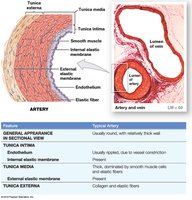
Differences Between Arteries and Veins
- Arteries have thicker walls and higher blood pressure. - Arteries have a small, round lumen; veins have a large, irregular lumen. - Arteries are more elastic; veins have valves to prevent backflow.
Classification of Arteries
Elastic Arteries (Conducting Arteries)
- Large vessels (e.g., pulmonary trunk, aorta). - Tunica media contains many elastic fibers, few muscle cells. - Elasticity evens out pulse force.
Muscular Arteries (Distribution Arteries)
- Medium-sized arteries; tunica media has many muscle cells.
Arterioles (Resistance Vessels)
- Small vessels; little or no tunica externa, thin or incomplete tunica media. 
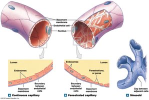
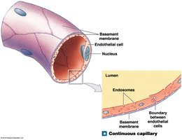
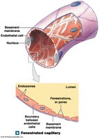
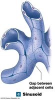
Capillaries: Structure and Function
Capillaries are the smallest vessels with thin walls, forming networks that permeate all active tissues. They are the site of exchange for nutrients, gases, and wastes between blood and interstitial fluid.
Capillary Structure
- Composed of an endothelial tube inside a thin basement membrane. - No tunica media or externa. - Diameter similar to a red blood cell.
Types of Capillaries
Type | Structure | Function | Location |
|---|---|---|---|
Continuous | Complete endothelial lining | Permits diffusion of water, small solutes, lipid-soluble materials; blocks blood cells and plasma proteins | All tissues except epithelia and cartilage; specialized forms in CNS and thymus (blood-brain barrier) |
Fenestrated | Pores in endothelial lining | Permits rapid exchange of water and larger solutes | Choroid plexus, endocrine organs, kidneys, intestinal tract |
Sinusoids | Gaps between adjacent endothelial cells | Permits free exchange of water and large plasma proteins | Liver, spleen, bone marrow, endocrine organs |

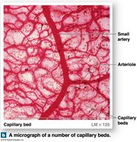
Capillary Beds and Blood Flow Regulation
- Capillary beds connect one arteriole and one venule. - Precapillary sphincters guard entrance to each capillary, opening and closing to regulate blood flow. - Thoroughfare channels provide direct connections between arterioles and venules. 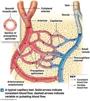
Collateral Circulation and Anastomoses
- Collaterals: Multiple arteries supply one capillary bed, allowing circulation if one artery is blocked. - Arterial anastomosis: Fusion of two collateral arteries. - Arteriovenous anastomoses: Direct connections between arterioles and venules, bypassing capillary beds.
Angiogenesis
- Formation of new blood vessels, stimulated by vascular endothelial growth factor (VEGF). - Occurs during embryonic development and in response to hypoxia.
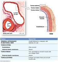
Structure and Function of Veins
Veins collect blood from capillaries and return it to the heart. Compared to arteries, veins have larger diameters, thinner walls, and lower blood pressure.
Types of Veins
- Venules: Very small veins collecting blood from capillaries. - Medium-sized veins: Thin tunica media, few smooth muscle cells, tunica externa with elastic fibers. - Large veins: All three tunica layers, thick tunica externa, thin tunica media.
Venous Valves
- Folds of tunica intima prevent backflow of blood. - Compression of veins pushes blood toward the heart. - Weak walls near valves can result in varicose veins or hemorrhoids. 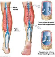
Distribution of Blood in the Cardiovascular System
- Heart, arteries, and capillaries contain 30–35% of blood volume. - Venous system contains 65–70% of blood volume; one-third is in large venous networks of the liver, bone marrow, and skin. 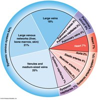
Capacitance of Blood Vessels
- Capacitance is the ability to stretch. - Veins stretch more than arteries and act as blood reservoirs. - Venoconstriction increases blood in arterial system and capillaries during blood loss.
Pressure and Resistance in the Cardiovascular System
Factors Influencing Blood Pressure
Blood flow equals cardiac output and is determined by pressure (P) and resistance (R).
Pressure Gradient
- Flow (F) is proportional to the pressure gradient (∆P) divided by resistance (R):
Types of Pressure
- Blood Pressure (BP): Arterial pressure (mm Hg). - Capillary Hydrostatic Pressure (CHP): Pressure within capillary beds. - Venous Pressure: Pressure in the venous system.
Total Peripheral Resistance
- Must overcome resistance of the entire cardiovascular system. - ∆P across systemic circuit ≈ 85 mm Hg.
Components of Resistance
- Vascular Resistance: Due to friction between blood and vessel walls; depends on vessel length and diameter. - Blood Viscosity: Resistance caused by molecules and suspended materials; whole blood viscosity ≈ 4x water. - Turbulence: Swirling action that disturbs smooth flow; occurs in heart chambers and great vessels. 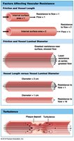
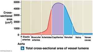
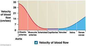
Cardiovascular Pressures and Blood Flow
- Vessel luminal diameters, total cross-sectional areas, pressures, and velocity of blood flow vary throughout the systemic circuit. 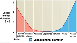
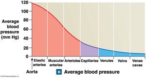
Arterial Blood Pressure
- Systolic Pressure: Peak arterial pressure during ventricular systole. - Diastolic Pressure: Minimum arterial pressure at end of ventricular diastole. - Pulse Pressure: Difference between systolic and diastolic pressure. - Mean Arterial Pressure (MAP):
Normal and Abnormal Blood Pressure
- Normal: 120/80 mm Hg. - Hypertension: >140/90 mm Hg. - Hypotension: <90/60 mm Hg.
Elastic Rebound
- Arterial walls stretch during systole and recoil during diastole, maintaining blood flow. 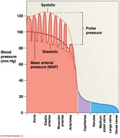
Venous Pressure and Return
- Venous pressure determines venous return to the heart. - Assisted by skeletal muscle compression and the respiratory pump.
Capillary Exchange
Capillary exchange is vital to homeostasis, allowing movement of materials across capillary walls by diffusion, filtration, and reabsorption.
Diffusion
- Movement of ions/molecules from high to low concentration. - Routes: Between endothelial cells, through pores, channels, or plasma membranes.
Filtration
- Driven by hydrostatic pressure; water and small solutes forced through capillary wall. 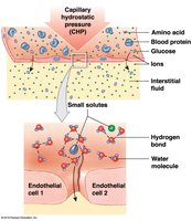
Reabsorption
- Result of osmosis; higher solute concentration leads to higher osmotic pressure. - Blood colloid osmotic pressure (BCOP) prevents osmosis, caused by suspended blood proteins.
Interplay Between Filtration and Reabsorption
- Ensures constant communication between plasma and interstitial fluid. - Accelerates distribution of nutrients, hormones, and gases. - Assists transport of insoluble lipids and tissue proteins. - Carries toxins and stimuli to lymphatic tissues.
Net Filtration Pressure (NFP)
- NFP is the difference between net hydrostatic and net osmotic pressure: 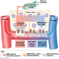
Capillary Dynamics
- Hemorrhaging reduces CHP and NFP, increasing reabsorption. - Dehydration increases BCOP, accelerating reabsorption. - If CHP rises or BCOP declines, fluid moves out of blood, causing edema.
Regulation of Blood Flow and Pressure in Tissues
Tissue Perfusion
- Blood flow through tissues delivers O2 and nutrients, removes CO2 and wastes. - Affected by cardiac output, peripheral resistance, and blood pressure.
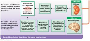
Cardiovascular Regulation Mechanisms
Vasomotion
- Contraction and relaxation cycle of precapillary sphincters, causing variable blood flow in capillary beds.
Control Mechanisms
- Autoregulation: Immediate, localized adjustments. - Neural Mechanisms: Rapid responses to changes at specific sites. - Endocrine Mechanisms: Direct long-term changes. 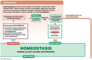
Autoregulation of Blood Flow
- Adjusted by peripheral resistance; precapillary sphincters constrict or dilate. - Local vasoconstrictors (e.g., endothelins, prostaglandins, thromboxanes) reduce blood flow. - Local vasodilators (e.g., low O2, high CO2, lactate, NO, histamine, elevated temperature) increase blood flow.
Neural Mechanisms
- Cardiovascular center in medulla oblongata: Cardioacceleratory center increases cardiac output; cardioinhibitory center reduces it. - Vasomotor center controls vasoconstriction (adrenergic nerves) and vasodilation (cholinergic nerves). - Vasomotor tone is produced by constant sympathetic vasoconstrictor action.
Reflex Control
- Baroreceptor reflexes respond to changes in blood pressure. - Chemoreceptor reflexes respond to changes in pH and dissolved gases. 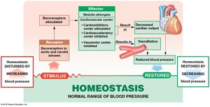
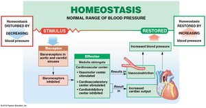
Baroreceptor Reflexes
- Stretch receptors in carotid sinuses, aortic sinuses, and right atrium monitor blood pressure. - Aortic reflex adjusts blood pressure and flow in systemic circuit. - When blood pressure rises: CV centers decrease cardiac output and cause peripheral vasodilation. - When blood pressure falls: CV centers increase cardiac output and cause peripheral vasoconstriction. - Bainbridge reflex responds to stretching of right atrium.
Chemoreceptor Reflexes
- Peripheral chemoreceptors in carotid and aortic bodies monitor blood. - Central chemoreceptors below medulla oblongata monitor cerebrospinal fluid. - Respond to changes in pH, O2, and CO2; coordinate cardiovascular and respiratory activities. 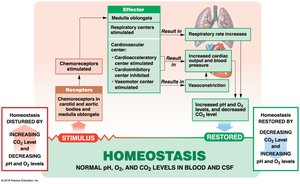
Endocrine Mechanisms
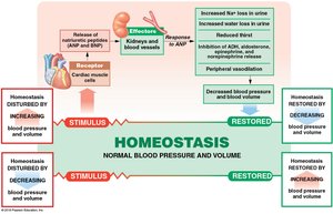
- Hormones have short-term and long-term effects. - Epinephrine and norepinephrine stimulate cardiac output and vasoconstriction. - Antidiuretic hormone (ADH) elevates blood pressure and reduces water loss. - Angiotensin II stimulates aldosterone, ADH, thirst, cardiac output, and vasoconstriction. - Erythropoietin (EPO) stimulates vasoconstriction and red blood cell production. - Natriuretic peptides (ANP, BNP) reduce blood volume and pressure. 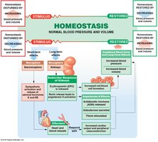
Cardiovascular Adaptation
Special Circulation
Some organs have unique mechanisms to control blood flow:
Brain
- Blood flow is top priority; cerebral vessels dilate when peripheral vessels constrict. - Cerebrovascular accident (stroke) results from blockage or rupture in a cerebral artery.
Heart
- Coronary arteries supply blood; demand increases with activity. - Lactic acid and low O2 dilate coronary vessels; epinephrine increases heart rate and strength. - Heart attack results from blockage of coronary blood flow.
Lungs
- Perfusion regulated by O2 levels in alveoli; high O2 dilates vessels, low O2 constricts them.
Cardiovascular Response to Exercise
Light Exercise
- Extensive vasodilation increases circulation. - Venous return increases with muscle contractions. - Cardiac output rises due to venous return and atrial stretching.
Heavy Exercise
- Activates sympathetic nervous system; cardiac output increases to maximum. - Blood flow is redirected to skeletal muscles, lungs, and heart; supply to brain is unaffected.
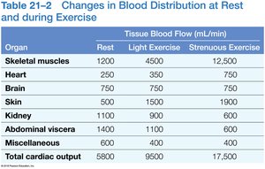
Organ | Rest | Light Exercise | Strenuous Exercise |
|---|---|---|---|
Skeletal muscles | 1200 | 4500 | 12500 |
Heart | 250 | 350 | 750 |
Brain | 750 | 750 | 750 |
Skin | 500 | 1500 | 750 |
Kidney | 1100 | 1100 | 600 |
Abdominal viscera | 1400 | 1100 | 600 |
Miscellaneous | 600 | 400 | 300 |
Total cardiac output | 5800 | 9500 | 17500 |
Cardiovascular Response to Hemorrhaging
- Short-term elevation of blood pressure: Carotid and aortic reflexes increase cardiac output and cause vasoconstriction. - Sympathetic nervous system further constricts arterioles; venoconstriction improves venous return. - Hormonal effects increase cardiac output and peripheral vasoconstriction. - Failure to restore blood pressure results in shock. - Long-term restoration: Recall of fluids, aldosterone and ADH promote fluid retention, thirst increases, EPO stimulates RBC production. 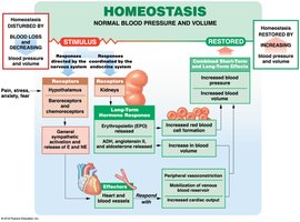
Major Arteries and Veins of the Systemic Circuit
Arteries of the Neck and Head
- Carotid sinus contains baroreceptors for blood pressure regulation. 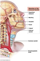
Arteries Supplying Abdominopelvic Organs
- Arterial blood supply to the liver is crucial for nutrient processing. 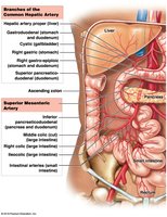
The Hepatic Portal System
- Connects two capillary beds; delivers nutrient-laden blood from digestive organs to the liver for processing. - Blood collects in hepatic veins and empties into the inferior vena cava. 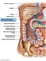
Summary of Learning Outcomes
Distinguish among types of blood vessels based on structure and function.
Describe factors influencing blood pressure and explain capillary exchange.
Explain control mechanisms regulating blood flow and pressure in tissues.
Identify principal blood vessels and functions of special circulation to brain, heart, and lungs; describe cardiovascular responses to exercise and hemorrhaging.
Describe pulmonary and systemic circuits; identify major arteries and veins.
Discuss differences between fetal and adult circulation; describe changes at birth.
Discuss effects of aging on the cardiovascular system and interactions with other organ systems.
