 Back
Blood Vessels and Circulation: Structure, Function, and Regulation
Study Guide - Smart Notes
 Tailored notes based on your materials, expanded with key definitions, examples, and context.
Blood Vessels: Overview and Classification
Types of Blood Vessels
The vascular system is composed of several types of blood vessels, each with distinct structures and functions. These include arteries, arterioles, capillaries, venules, and veins.
Arteries: Carry blood away from the heart under high pressure.
Arterioles: Small branches of arteries that regulate blood flow into capillaries.
Capillaries: Microscopic vessels where exchange of gases, nutrients, and wastes occurs between blood and tissues.
Venules: Small vessels that collect blood from capillaries and transport it to veins.
Veins: Return blood to the heart under lower pressure and often contain valves to prevent backflow.
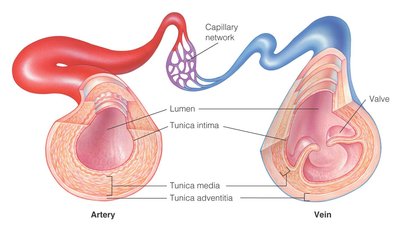
Structure of Blood Vessel Walls
Three Layers (Tunics) of Blood Vessels
Both arteries and veins have three main layers, but their thickness and composition vary:
Tunica Externa (Adventitia): Outermost connective tissue layer, provides structural support and attachment for nerves and lymphatics.
Tunica Media: Middle layer of smooth muscle and elastic fibers, responsible for vasoconstriction and vasodilation.
Tunica Interna (Intima): Innermost layer of squamous epithelium (endothelium), minimizes friction between blood and vessel wall.
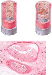
Arteries and Arterioles
Functional Characteristics
Arteries: Act as pressure reservoirs, maintaining blood flow during ventricular relaxation. Large arteries are elastic, while smaller ones are muscular and distribute blood to organs.
Arterioles: Known as resistance vessels, they regulate blood flow into capillaries by altering their diameter through contraction or relaxation of the tunica media. This process is called vasoconstriction (narrowing) and vasodilation (widening).
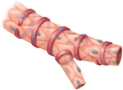
Capillaries
Structure and Function
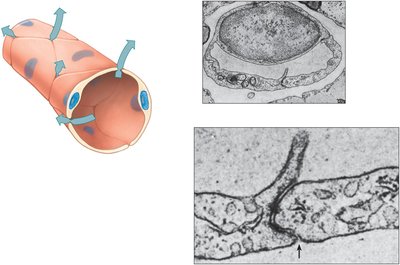
Capillaries are the smallest blood vessels, consisting of a single layer of endothelial cells. They connect arterioles to venules and are the primary sites for exchange of gases, nutrients, and waste products between blood and tissues.
Precapillary sphincters regulate blood flow into capillary beds.
Capillaries are semi-permeable, allowing selective exchange of substances.
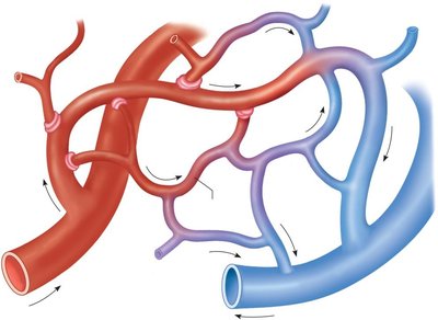
Capillary Permeability
Capillary walls allow for the movement of fluids and small solutes. The structure of endothelial cells and the presence of intercellular clefts or fenestrations determine permeability.
Venules and Veins
Structure and Function
Venules: Collect blood from capillaries; have thinner walls and less smooth muscle than arterioles.
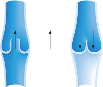
Veins: Have thinner walls than arteries, larger lumens, and often contain valves to prevent backflow. They function as volume reservoirs, holding the majority of the body's blood at rest.
Venous Return Mechanisms
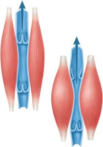
Skeletal muscle contraction: Compresses veins, pushing blood toward the heart.
Respiratory movements: Changes in thoracic pressure during breathing aid venous return.
Venoconstriction: Contraction of smooth muscle in vein walls increases venous return.
Valves: Prevent backflow of blood in veins, especially in limbs.
Blood Pressure and Its Regulation
Blood Pressure Definitions
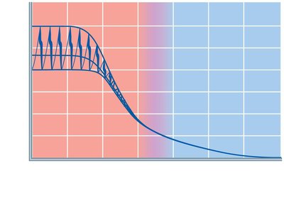
Blood pressure (BP): The force exerted by circulating blood on the walls of blood vessels.
Systolic pressure: Maximum pressure during ventricular contraction.
Diastolic pressure: Minimum pressure during ventricular relaxation.
Mean Arterial Pressure (MAP): Average pressure in arteries during one cardiac cycle.
MAP Calculation:
Factors Affecting Blood Pressure
Blood volume: Increased volume raises BP.
Heart rate and stroke volume: Increased cardiac output raises BP.
Blood viscosity: Thicker blood increases resistance and BP.
Peripheral resistance: Narrower vessels increase resistance and BP.
Baroreceptor Reflex
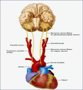
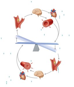
Baroreceptors are stretch-sensitive mechanoreceptors located in the aortic arch and carotid sinuses. They detect changes in blood pressure and initiate reflexes to maintain homeostasis.
Increased BP stimulates baroreceptors, leading to decreased heart rate and vasodilation.
Decreased BP reduces baroreceptor firing, increasing heart rate and vasoconstriction.
Neural and Hormonal Regulation
Neural: Vasomotor center in the medulla oblongata adjusts vessel diameter via sympathetic and parasympathetic pathways.
Hormonal:
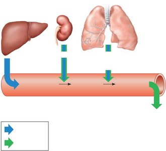
Adrenal medulla hormones (epinephrine, norepinephrine): Increase heart rate, cardiac output, and vasoconstriction.
Renin-angiotensin-aldosterone system (RAAS): Increases BP via vasoconstriction and increased blood volume.
Antidiuretic hormone (ADH): Promotes water reabsorption in kidneys, increasing blood volume and BP.
Atrial natriuretic peptide (ANP): Decreases BP by increasing sodium and water excretion.
Systemic Circulation: Major Arteries and Veins
Major Arteries of the Systemic Circuit
Aorta: Main artery leaving the left ventricle, branches into the brachiocephalic, left common carotid, and left subclavian arteries.
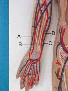
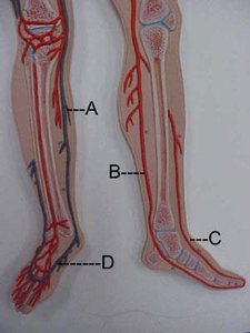
Subclavian, axillary, brachial, radial, and ulnar arteries: Supply the upper limbs.
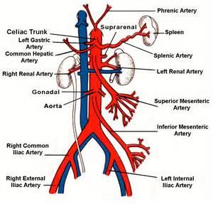
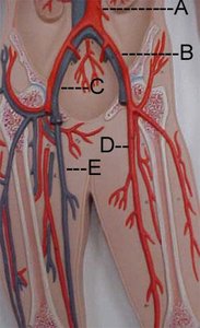
Thoracic and abdominal aorta: Supply the trunk and lower limbs via branches such as the celiac trunk, mesenteric, renal, and iliac arteries.
Major Veins of the Systemic Circuit
Superficial and deep veins often parallel arteries but have more variability in naming and structure.
Superior vena cava (SVC): Returns blood from the upper body to the right atrium.
Inferior vena cava (IVC): Returns blood from the lower body to the right atrium.
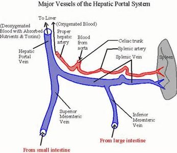
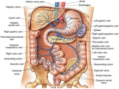
Hepatic portal vein: Drains blood from digestive organs to the liver for processing before entering systemic circulation.
Vessel Dynamics and Blood Flow Equations
Key Equations
Cardiac Output (CO): The volume of blood ejected by the heart per minute. Where HR is heart rate and SV is stroke volume.
Blood Pressure (BP): Determined by cardiac output and peripheral resistance. Where R is resistance.
Factors affecting resistance:
Vessel radius (smaller radius increases resistance)
Vessel length (longer vessels increase resistance)
Blood viscosity (thicker blood increases resistance)
Additional info: Blood pressure is primarily regulated by the sympathetic nervous system, while heart rate is mainly under parasympathetic control.
