 Back
Blood Vessels and Circulation: Structure, Function, and Regulation
Study Guide - Smart Notes
 Tailored notes based on your materials, expanded with key definitions, examples, and context.
Blood Vessels: Overview and Classification
Introduction to Blood Vessels
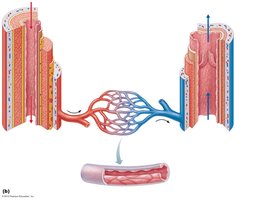
The blood vessels form a dynamic delivery system that begins and ends at the heart, ensuring the transport of blood throughout the body. The three major types of blood vessels are arteries, capillaries, and veins.
Arteries: Carry blood away from the heart; typically oxygenated except in pulmonary circulation and fetal umbilical vessels.
Capillaries: Serve as sites of exchange between blood and tissues, directly serving cellular needs.
Veins: Return blood toward the heart.
Relationship of Blood Vessels and Lymphatics
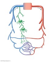
Blood vessels are closely associated with the lymphatic system, which helps maintain fluid balance and immune function. The arterial and venous systems are interconnected with lymphatic vessels and nodes.
Structure of Blood Vessel Walls
Layers of Vessel Walls
Most blood vessels (except capillaries) have three layers:
Tunica intima: Innermost layer, composed of endothelium and a subendothelial layer; may include an internal elastic membrane.
Tunica media: Middle layer, primarily smooth muscle and elastic fibers; responsible for vasoconstriction and vasodilation.
Tunica externa: Outermost layer, mainly collagen fibers; contains nerves and, in larger vessels, vasa vasorum (small vessels that supply the vessel wall).
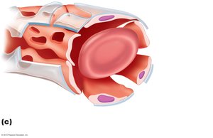
Capillaries consist only of endothelium and a basement membrane, facilitating exchange.
Types of Capillaries
Continuous Capillaries
Continuous capillaries are the least permeable and most common, found in skin and muscle. They have uninterrupted endothelial linings with tight junctions and intercellular clefts for limited passage of fluids and small solutes.
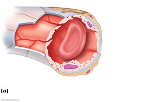
Fenestrated Capillaries
Fenestrated capillaries have pores (fenestrations) that increase permeability. They are found in areas of active absorption or filtration, such as the kidneys and small intestine.
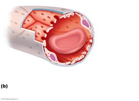
Sinusoid Capillaries
Sinusoid capillaries are the most permeable, with large gaps and an incomplete basement membrane. They are present in the liver, bone marrow, and spleen, allowing passage of large molecules and cells.
Capillary Beds and Microcirculation
Capillary Bed Structure and Flow Regulation
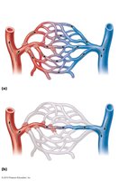
Capillary beds consist of a vascular shunt (metarteriole and thoroughfare channel) and true capillaries. Precapillary sphincters regulate blood flow into true capillaries, directing flow according to tissue needs.
Distribution of Blood Volume
Relative Proportion of Blood in the Vascular System
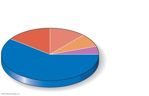
Blood volume is unevenly distributed in the cardiovascular system. The majority is found in systemic veins and venules, which act as blood reservoirs.
Location | Percentage of Blood Volume |
|---|---|
Pulmonary blood vessels | 12% |
Systemic arteries and arterioles | 15% |
Heart | 8% |
Capillaries | 5% |
Systemic veins and venules | 60% |
Physiology of Circulation
Key Terms and Concepts
Blood flow: Volume of blood flowing through a vessel, organ, or the entire circulation per unit time (ml/min). For the entire vascular system, it equals cardiac output (CO).
Blood pressure (BP): Force per unit area exerted on a vessel wall by the blood, measured in mm Hg. Systemic arterial BP is typically measured in large arteries near the heart.
Resistance (peripheral resistance): Opposition to blood flow due to friction between blood and vessel walls. Major sources include blood viscosity, vessel length, and vessel diameter (the latter has the greatest influence).
Relationship Between Flow, Pressure, and Resistance
Blood flow (F) is directly proportional to the pressure gradient (ΔP) and inversely proportional to resistance (R):
Resistance is most easily altered by changing vessel diameter (vasoconstriction increases resistance, vasodilation decreases it).
Blood Pressure Changes in the Systemic Circulation
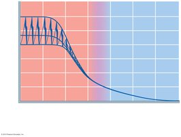
Blood pressure is highest in the aorta and declines throughout the pathway, reaching its lowest point in the venae cavae. The steepest drop occurs in the arterioles.
Arterial Blood Pressure
Systolic pressure: Pressure during ventricular contraction (average 120 mm Hg in adults).
Diastolic pressure: Lowest aortic pressure during relaxation.
Pulse pressure: Difference between systolic and diastolic pressure.
Mean arterial pressure (MAP): Average pressure propelling blood to tissues. Calculated as: Example: For BP 120/80, MAP = 93 mm Hg.
Venous Return and Its Regulation
Factors Aiding Venous Return
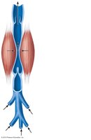
Muscular pump: Skeletal muscle contractions "milk" blood toward the heart; valves prevent backflow.
Respiratory pump: Pressure changes during breathing move blood toward the heart by compressing abdominal veins and expanding thoracic veins.
Venoconstriction: Sympathetic stimulation reduces vein diameter, pushing blood toward the heart.
Control of Blood Pressure
Short-Term Neural and Hormonal Controls
Neural controls operate via reflex arcs involving baroreceptors, the cardiovascular center of the medulla, vasomotor fibers, and sometimes chemoreceptors and higher brain centers.
Chemoreceptor reflexes respond to increased CO2, decreased pH, or low O2, increasing BP by stimulating the heart and vasoconstriction.
Higher brain centers (hypothalamus, cortex) can modify BP during stress, exercise, or temperature changes.
Hormonal controls:
Epinephrine, norepinephrine, angiotensin II, and high ADH increase BP.
Atrial natriuretic peptide decreases BP by reducing blood volume.
Long-Term Renal Regulation
Direct mechanism: Increased BP or blood volume leads to increased urine output, reducing BP; decreased BP causes water conservation.
Indirect mechanism (Renin-Angiotensin-Aldosterone): Low BP triggers renin release, leading to angiotensin II formation, which increases BP via vasoconstriction and aldosterone-mediated water retention.
Velocity of Blood Flow
Changes in Blood Flow Velocity
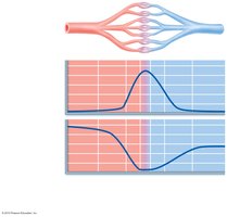
Blood flow velocity is inversely related to the total cross-sectional area of vessels. It is fastest in the aorta, slowest in capillaries (allowing exchange), and increases again in veins.
Autoregulation of Blood Flow
Intrinsic Regulation Mechanisms
Metabolic controls: Vasodilation in response to low O2, high temperature, or metabolic byproducts (H+, K+, adenosine, prostaglandins).
Myogenic controls: Vascular smooth muscle responds to stretch; increased pressure causes vasoconstriction, reduced stretch causes vasodilation.
Long-term autoregulation: Involves angiogenesis (growth of new vessels) when short-term mechanisms are insufficient.
Capillary Exchange and Bulk Flow
Mechanisms of Capillary Exchange
Diffusion: O2 and nutrients move from blood to tissues; CO2 and wastes move from tissues to blood.
Lipid-soluble substances diffuse through endothelial membranes; water-soluble substances pass through clefts and fenestrations; large molecules use vesicular transport.
Bulk Flow: Fluid Movements Across Capillary Walls
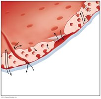
Bulk flow determines fluid distribution between blood and interstitial space. Fluid leaves capillaries at the arterial end and is reabsorbed at the venous end. The direction and amount of flow depend on hydrostatic and colloid osmotic pressures.
Hydrostatic and Osmotic Pressures
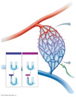
Capillary hydrostatic pressure (HPc): Pushes fluid out of capillaries; higher at arterial end (35 mm Hg) than venous end (17 mm Hg).
Interstitial fluid hydrostatic pressure (HPif): Usually zero due to lymphatic drainage.
Capillary colloid osmotic pressure (OPc): Pulls fluid into capillaries (~26 mm Hg), due to plasma proteins.
Interstitial fluid osmotic pressure (OPif): Low (~1 mm Hg).
Net Filtration Pressure (NFP)
NFP determines the direction of fluid movement:
At the arterial end: mm Hg (net outward pressure; filtration).
At the venous end: mm Hg (net inward pressure; reabsorption).
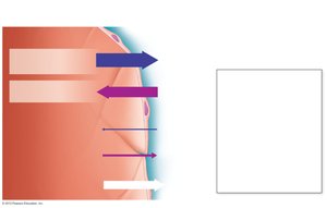
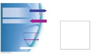
Summary: Bulk flow across capillary walls maintains fluid balance and the interstitial environment. About 20 L of fluid is filtered out daily, with 17 L reabsorbed and 3 L returned via the lymphatic system.
