 Back
Blood Vessels and Circulation: Structure, Function, and Physiology
Study Guide - Smart Notes
 Tailored notes based on your materials, expanded with key definitions, examples, and context.
Blood Vessels: Overview
Introduction to Blood Vessels
Blood vessels form a closed vascular system that transports blood throughout the body, ensuring the delivery of oxygen and nutrients to tissues and the removal of waste products. The three main types of blood vessels are arteries, veins, and capillaries, each with distinct structural and functional characteristics.
Arteries and arterioles: Carry blood away from the heart.
Capillaries: Facilitate exchange of substances between blood and tissues.
Venules and veins: Return blood toward the heart.
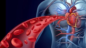
Anatomy of Blood Vessels
General Structure of Blood Vessel Walls
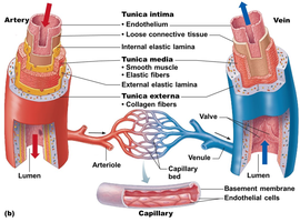
Except for capillaries, blood vessels have three distinct layers, or tunics, that provide structural support and regulate vessel function:
Tunica intima: Innermost layer; composed of endothelium that reduces friction for smooth blood flow.
Tunica media: Middle layer; consists of smooth muscle and elastic fibers, regulated by the sympathetic nervous system to control vessel diameter.
Tunica externa: Outermost layer; mainly fibrous connective tissue that protects and supports the vessel.
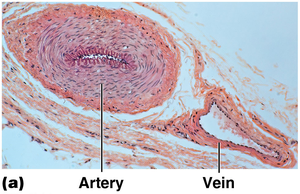
Microscopic Anatomy: Arteries vs. Veins
Arteries, veins, and capillaries differ in their wall structure and function:
Arteries: Thick, strong, and elastic tunica media to withstand high pressure.
Veins: Thinner tunica media, larger lumen, and valves to prevent backflow; operate under lower pressure.
Capillaries: Only a single layer of endothelium (tunica intima) to facilitate exchange.
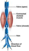
Venous Return Mechanisms
Veins rely on several mechanisms to return blood to the heart, especially from the lower body:
Valves: Prevent backflow of blood.
Skeletal muscle pump: Muscle contractions "milk" blood toward the heart.
Capillary Beds and Microcirculation
Structure and Function of Capillary Beds
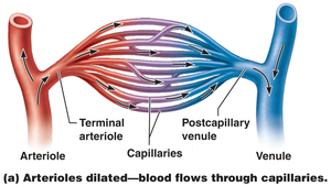
Capillary beds are networks of capillaries where exchange between blood and tissues occurs. Blood flow through these beds is called microcirculation.
Blood flows from terminal arterioles into capillary beds and then into postcapillary venules.
Capillaries are one cell layer thick, allowing efficient exchange of gases, nutrients, and wastes.
Regulation of Capillary Blood Flow
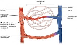
Specialized structures called precapillary sphincters regulate blood flow through capillary beds:
Precapillary sphincters: Rings of smooth muscle that open or close to control blood flow into capillaries.
Vascular shunt: Directly connects arteriole and venule, bypassing the capillary bed when sphincters are closed.
Effects of Arteriole Dilation and Constriction
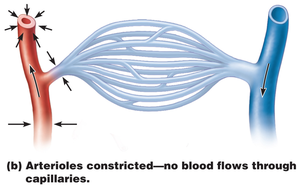
Blood flow through capillaries is influenced by the state of arterioles:
Dilated arterioles: Blood flows freely through capillaries, allowing exchange.
Constricted arterioles: Blood bypasses capillaries, reducing exchange in that region.
Gross Anatomy of Major Blood Vessels
Major Arteries of Systemic Circulation
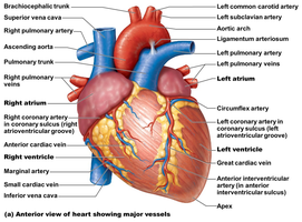
The aorta is the largest artery, originating from the left ventricle and branching into major arteries that supply the body:
Ascending aorta → Aortic arch → Thoracic aorta → Abdominal aorta
Branches supply the heart (coronary arteries), head, neck, upper limbs, thorax, abdomen, pelvis, and lower limbs.
Physiology of Circulation
Vital Signs and Blood Pressure
Vital signs include arterial pulse, blood pressure, respiratory rate, and body temperature. Blood pressure is the force exerted by blood against vessel walls and is essential for maintaining blood flow.
Arterial pulse: Expansion and recoil of arteries with each heartbeat; normal rate is 70–76 beats per minute at rest.
Blood pressure: Expressed as systolic/diastolic (e.g., 120/80 mm Hg); measured indirectly using the auscultatory method, usually at the brachial artery.
Blood Pressure Regulation
Blood pressure is determined by cardiac output (CO) and peripheral resistance (PR):
Cardiac output (CO): Volume of blood pumped by the heart per minute.
Peripheral resistance (PR): Friction encountered by blood as it flows through vessels.
Formula:
Neural factors: Sympathetic nervous system increases blood pressure via vasoconstriction; parasympathetic has little effect.
Renal factors: Kidneys regulate blood volume and pressure via water excretion or retention and the renin-angiotensin-aldosterone system.
Other factors: Temperature, chemicals (e.g., epinephrine), and diet can influence blood pressure.
Capillary Exchange and Fluid Movement
Exchange of gases, nutrients, and wastes occurs across capillary walls by diffusion, filtration, and vesicular transport. Fluid movement is governed by the balance between blood pressure and osmotic pressure:
At the arterial end: Blood pressure exceeds osmotic pressure, causing fluid to leave the capillary.
At the venous end: Osmotic pressure exceeds blood pressure, drawing fluid back into the capillary.
Developmental Aspects of the Cardiovascular System
Fetal Circulation
In the fetus, special adaptations allow blood to bypass the lungs and liver, which are not fully functional before birth:
Foramen ovale: Shunts blood from right atrium to left atrium.
Ductus arteriosus: Connects pulmonary trunk to aorta.
Ductus venosus: Bypasses the liver, directing blood to the inferior vena cava.
Umbilical vein and arteries: Carry oxygenated blood to the fetus and deoxygenated blood to the placenta, respectively.
Age-Related Changes
With aging, the cardiovascular system undergoes several changes:
Weakening of venous valves (leading to varicose veins)
Progressive arteriosclerosis (hardening of arteries)
Increased risk of hypertension and coronary artery disease due to loss of vessel elasticity and fatty deposits
