 Back
BackBlood Vessels and Circulation: Structure, Function, and Regulation
Study Guide - Smart Notes
 Tailored notes based on your materials, expanded with key definitions, examples, and context.
Blood Vessels and Circulation
Structure of Vessel Walls
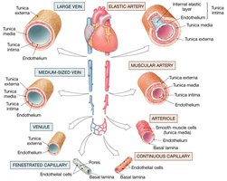
The walls of arteries and veins are composed of three distinct layers, each contributing to the vessel's function and integrity.
Tunica intima: The innermost layer, consisting of endothelium and a subendothelial layer.
Tunica media: The middle layer, primarily composed of smooth muscle and elastic fibers, responsible for vasoconstriction and vasodilation.
Tunica externa: The outermost layer, made of connective tissue that provides structural support and protection.
Differences Between Arteries and Veins
Arteries and veins differ in structure and function to accommodate their roles in the circulatory system.
Arteries have thicker walls, more smooth muscle, and elastic fibers, making them more resilient to pressure changes.
Veins have thinner walls and less smooth muscle, making them more compliant but less resilient.
Types of Arteries
Arteries are classified based on their size and function:
Elastic arteries (conducting arteries): Large arteries (up to 1 inch in diameter) that transport blood away from the heart and help dampen pressure fluctuations.
Muscular arteries (distribution arteries): Medium-sized arteries (about 4 mm in diameter) that distribute blood to specific organs.
Arterioles: Small arteries (internal diameter of 30 μm or less) that regulate blood flow into capillary beds.
Capillaries
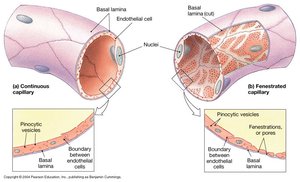
Capillaries are the smallest blood vessels, consisting of an endothelial tube within a basal lamina. They facilitate the exchange of gases, nutrients, and wastes between blood and tissues.
Form networks that surround muscle fibers and radiate through connective tissue.
Two main types: Continuous capillaries (with uninterrupted endothelial lining) and fenestrated capillaries (with pores for increased permeability).
Sinusoids: Flattened, fenestrated capillaries found in organs like the liver and spleen.
Capillary Beds
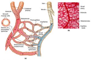
Capillary beds are interconnected networks of vessels that regulate blood flow to tissues.
Components include collateral arteries, metarterioles, arteriovenous anastomoses, capillaries, and venules.
Precapillary sphincters control the flow of blood into capillary beds.
Veins
Veins collect blood from tissues and return it to the heart. They are classified by size:
Venules: Smallest veins, collecting blood from capillaries.
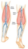
Medium-sized veins: Contain valves to prevent backflow.
Large veins: Include the superior and inferior vena cava.
Venous Valves
Venules and medium-sized veins contain valves that prevent the backflow of blood, especially in the limbs, ensuring unidirectional flow toward the heart.
Distribution of Blood
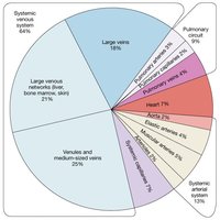
Blood volume is unevenly distributed within the cardiovascular system. Veins act as capacitance vessels, accommodating large volumes of blood with little change in pressure.
Venoconstriction helps maintain blood volume during blood loss or redistribution.
Capacitance is the relationship between blood volume and pressure.
Circulatory Pressure
Blood flow is determined by the pressure gradient and resistance within the vessels.
Flow is proportional to the pressure difference ().
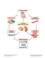
Circulatory pressure is divided into blood pressure (BP), capillary hydrostatic pressure (CHP), and venous pressure.
Cardiovascular Physiology Overview
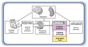
The cardiovascular system maintains blood flow and pressure through a complex interplay of cardiac output, vessel resistance, and regulatory mechanisms.
Resistance (R)
Resistance opposes blood flow and is primarily determined by the arterial system (peripheral resistance).
Factors affecting resistance include vessel diameter, blood viscosity, and total vessel length.
For blood to flow, the pressure gradient must overcome total peripheral resistance.
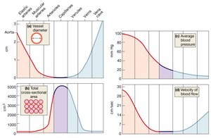
Relationships Among Vessel Diameter, Cross-sectional Area, Blood Pressure, and Blood Viscosity
These factors collectively influence blood flow and pressure throughout the vascular system.
Arterial Blood Pressure
Arterial blood pressure maintains blood flow through capillary beds and fluctuates with the cardiac cycle.
Rises during ventricular systole and falls during diastole.
Pulse pressure is the difference between systolic and diastolic pressures.
Mean arterial pressure (MAP) is calculated as:
Capillary Exchange
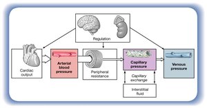
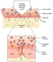
Capillary exchange is the movement of water and solutes between blood and interstitial fluid, essential for nutrient delivery and waste removal.
Processes include diffusion, filtration (driven by hydrostatic pressure), and reabsorption (driven by osmotic pressure).
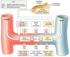
Forces involved: capillary hydrostatic pressure (CHP), blood colloid osmotic pressure (BCOP), interstitial fluid colloid osmotic pressure (ICOP), and interstitial fluid hydrostatic pressure (IHP).
Forces Acting Across Capillary Walls
Water movement across capillary walls is determined by the balance of hydrostatic and osmotic pressures.
Net hydrostatic pressure:
Net colloid osmotic pressure:
Net filtration pressure (NFP) determines the direction and magnitude of fluid movement.
Venous Pressure and Venous Return
Venous return is assisted by muscular compression and the respiratory pump, ensuring efficient return of blood to the heart.
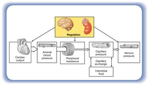
Cardiovascular Regulation
The cardiovascular system uses multiple mechanisms to maintain adequate blood flow and pressure:
Autoregulation: Local mechanisms that adjust blood flow within tissues.
Neural mechanisms: Adjust cardiac output and peripheral resistance via the autonomic nervous system.
Endocrine mechanisms: Hormones that enhance short-term and direct long-term changes in blood pressure and volume.
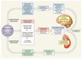
Autoregulation of Blood Flow
Local vasodilators accelerate blood flow by relaxing smooth muscle in response to:
Decreased tissue O2 or increased CO2
Generation of lactic acid
Release of nitric oxide (NO)
Rising K+ or H+ concentrations
Local inflammation (histamine, NO)
Elevated temperature
Neural Mechanisms
Neural mechanisms adjust cardiac output and peripheral resistance to maintain blood flow to vital organs.
Medullary centers include cardiac and vasomotor centers.
Vasoconstriction is mediated by norepinephrine (NE); vasodilation by nitric oxide (NO).
Reflex Control of Cardiovascular Function
Baroreceptor and chemoreceptor reflexes monitor and adjust cardiovascular function:
Baroreceptors: Monitor stretch and blood pressure in the carotid and aortic sinuses.
Chemoreceptors: Monitor CO2, O2, and pH levels in blood.
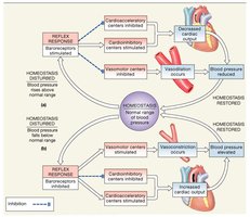
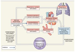
Hormonal Regulation of Cardiovascular Function
Several hormones regulate blood pressure and volume:
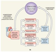
Antidiuretic hormone (ADH): Released in response to decreased blood volume, promotes water retention.
Angiotensin II: Produced in response to low blood pressure, causes vasoconstriction and stimulates aldosterone release.
Aldosterone: Promotes sodium and water retention, increasing blood volume.
Erythropoietin (EPO): Stimulates red blood cell production if O2 levels are low.
Natriuretic peptides: Released in response to excessive atrial stretch, promote sodium and water excretion.
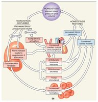
Exercise and the Cardiovascular System
Exercise induces significant changes in cardiovascular function:
Light exercise: Extensive vasodilation, increased venous return, and a rise in cardiac output.
Heavy exercise: Increased blood flow to skeletal muscles, restriction of flow to nonessential organs.
Cardiovascular Response to Hemorrhaging
The body responds to blood loss through short-term and long-term mechanisms:
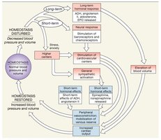
Short-term: Baroreceptor and chemoreceptor reflexes increase cardiac output and peripheral vasoconstriction; sympathetic activation elevates blood pressure; hormones like epinephrine, norepinephrine, and ADH are released.
Long-term: Fluid recall from interstitial spaces, increased thirst, aldosterone and ADH promote fluid retention, and erythropoietin increases red blood cell production.
Additional info: This guide integrates foundational concepts from Chapter 21 of Anatomy & Physiology, focusing on the structure and function of blood vessels, mechanisms of blood flow regulation, and physiological responses to changes in blood pressure and volume.
