 Back
Blood Vessels and Lymphatic System: Structure, Function, and Clinical Correlations
Study Guide - Smart Notes
 Tailored notes based on your materials, expanded with key definitions, examples, and context.
Blood Vessels
Types of Blood Vessels
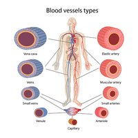
Blood vessels are essential components of the circulatory system, responsible for transporting blood throughout the body. They are classified into arteries, veins, and capillaries based on their structure and function.
Arteries: Carry blood away from the heart, typically under higher pressure. Can be oxygenated (systemic circulation) or deoxygenated (pulmonary circulation).
Veins: Carry blood toward the heart, under lower pressure. More likely to collapse under extremely low pressure. Can be oxygenated or deoxygenated.
Capillaries: Have the thinnest walls and serve as the site of exchange of gases and nutrients between blood and tissues.
Vessel Wall Tunics
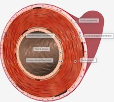
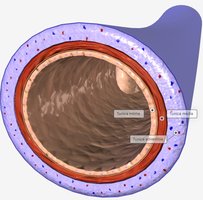
The walls of blood vessels are composed of three distinct layers, known as tunics, each with specific structural and functional roles.
Tunica intima: The innermost layer, consisting of endothelial cells that provide a smooth lining for blood flow.
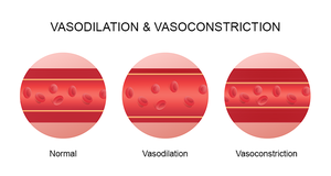
Tunica media: Composed of smooth muscle cells; thicker in arteries. Regulates vessel diameter through vasoconstriction and vasodilation.
Tunica externa: The outermost protective layer, stabilizing the vessel and composed mainly of connective tissue.
Lumen: The hollow space within the vessel; larger in veins.
Classification of Arteries
Arteries are further classified based on their function and structure:
Elastic arteries: Largest arteries, able to stretch and recoil to accommodate blood flow.
Distributing (muscular) arteries: Regulate distribution of blood to various organs.
Resistance arteries (arterioles): Regulate blood flow into capillaries and blood pressure, especially during activities like exercise.
Clinical Correlations
Aneurysm: Localized dilation or bulging of a blood vessel wall, which can lead to rupture.
Atherosclerosis: Accumulation of fatty plaques within arterial walls, leading to narrowing and reduced blood flow.
Arteriosclerosis: Hardening of the arteries, often associated with aging.
Arterial Sense Organs
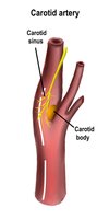
Specialized structures in arteries monitor blood pressure and chemical composition:
Carotid sinuses: Baroreceptors that monitor blood pressure.
Carotid bodies: Chemoreceptors that monitor pH, O2, and CO2 levels.
Aortic bodies: Chemoreceptors that monitor pH, O2, and CO2 levels.
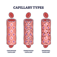
Capillary Types
Capillaries are classified based on their permeability:
Continuous capillaries: Have uninterrupted endothelial lining; most common type.
Fenestrated capillaries: Possess small pores that allow fluid and solutes to pass through.
Sinusoidal capillaries: Have large holes, permitting passage of proteins and cells.
Vein Classification
Veins are categorized based on size and function:
Postcapillary venules: Collect blood from capillaries.
Muscular veins: Medium-sized veins.
Venous sinuses: Large veins with thin walls.
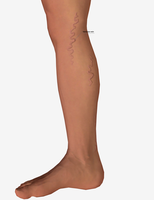
Clinical Correlation: Varicose Veins
Varicose veins: Result from dysfunctional valves, often due to age, prolonged increased abdominal pressure, prolonged sitting or standing, obesity, or pregnancy.
Hemorrhoids: Varicose veins of the anal canal.
Circulatory Route
The general pathway of blood flow is:
Heart → artery → arteriole → capillary → postcapillary venule → vein → heart
Anastomoses: Alternative pathways for blood flow.
Major Arteries and Veins
The Aorta and Its Major Branches
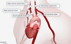
The aorta is the main artery with the highest pressure, branching into several major arteries:
Ascending aorta: Gives rise to left and right coronary arteries (supplying the heart muscle).
Aortic arch: Branches into brachiocephalic trunk, right common carotid, right subclavian, left common carotid, and left subclavian arteries.
Descending aorta: Supplies lower regions of the body.
Arteries and Veins of the Head and Neck
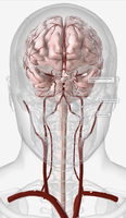
External carotid artery: Supplies superficial structures of the head.
Internal carotid artery: Supplies the brain via the Circle of Willis.
Vertebral arteries: Also supply the brain.
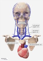
External jugular vein: Drains superficial structures of the head.
Internal jugular vein: Drains the brain.
Vertebral veins: Drain the brain.
Arteries and Veins of the Upper Limbs
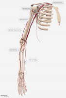
Arteries: Right subclavian, axillary, brachial (site for blood pressure cuff), radial, and ulnar (radial pulse).
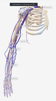
Veins: Deep veins (radial, ulnar, brachial, axillary, subclavian) and superficial veins (median cubital, cephalic, basilic).
Arteries and Veins of the Thoracic Cavity
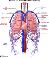
Thoracic aorta: Supplies visceral branches (pericardial, bronchial, esophageal, mediastinal) and parietal branches (posterior intercostal, subcostal, superior phrenic).
Azygos vein: Drains thoracic wall.
Inferior vena cava: Drains lower body.
Superior vena cava: Drains upper body.
Arteries and Veins of the Abdominopelvic Region
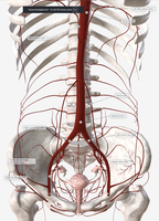
Abdominal aorta: Branches into inferior phrenic, celiac trunk (gastric, splenic, common hepatic), superior mesenteric, renal, inferior mesenteric, common iliac (internal and external branches).
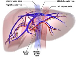
Hepatic portal vein: Receives blood from splenic, gastric, superior and inferior mesenteric veins.
Arteries and Veins of the Lower Limb
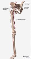
Arteries: Common iliac, external iliac, deep femoral, femoral, popliteal, anterior/posterior tibial, fibular.
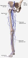
Veins: Superficial (small saphenous, great saphenous), deep (popliteal, femoral, deep femoral, external iliac, common iliac).
Blood Flow, Pressure, and Regulation
Flow and Perfusion
Blood flow refers to the volume of blood moving through vessels, while perfusion is the flow per mass or volume of tissue.
Blood Pressure
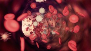
Blood pressure is the force exerted by blood against vessel walls, highest in the aorta. It is measured as systolic (during heart contraction) over diastolic (during heart relaxation).
Pulse Pressure: Difference between systolic and diastolic pressure.
Mean Arterial Pressure (MAP):
Peripheral Resistance
Peripheral resistance is influenced by blood viscosity, vessel length, and vessel diameter. Vasoconstriction increases resistance, while vasodilation decreases it.
Regulation of Blood Flow
Local Control: Autoregulation via vasoactive chemicals.
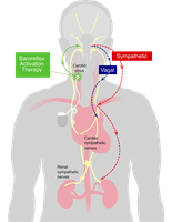
Neuronal Control: Baroreflex, chemoreflex, medullary ischemic reflex.
Hormonal Control: Angiotensin II, aldosterone, natriuretic peptides, antidiuretic hormone, catecholamines (epinephrine/norepinephrine).
Capillaries and Fluid Exchange
Capillaries facilitate exchange of fluids, nutrients, and waste via diffusion, transcytosis, filtration, and reabsorption.
Capillary hydrostatic pressure: Drives fluid out of capillaries.
Capillary osmotic pressure: Albumin drives fluid into capillaries.
Venous Return Mechanisms
Venous valves: Prevent backflow.
Skeletal muscle contractions: Pump blood toward the heart.
Respiratory pump: Assists venous return.
Gravity: Affects blood flow in upright posture.
Homeostatic Imbalances
Thrombus: Blood clot within a vessel.
Transient ischemic attacks: Temporary reduction in blood flow to the brain.
Cerebrovascular accidents: Stroke.
Circulatory shock: Includes cardiogenic, low venous return, hypovolemic, obstructed venous return, venous pooling, neurogenic shock.
Edema: Caused by increased capillary filtration, reduced reabsorption, or obstructed lymphatic drainage.
Lymphatic System
Functions of the Lymphatic System
The lymphatic system is vital for fluid recovery, immunity, and lipid absorption.
Fluid Recovery: Absorbs excess interstitial fluid and returns it to the bloodstream.
Immunity: Immune surveillance via lymph nodes, tonsils, and spleen.
Lipid Absorption: Lacteals absorb dietary lipids in the digestive system.
Lymph
Lymph: Colorless, clear, or pale yellow fluid recovered from tissues. Contains fewer proteins than plasma, may contain pathogens and white blood cells.
Lymphatic Vessels
Lymphatic vessels transport lymph under very low pressure and lack a central pump. They rely on skeletal muscle contractions, respiratory movements, and valves to prevent backflow.
Unique feature: Closed/blind-ended capillaries.
Flow pathway: Lymphatic capillaries → collecting vessels → lymph nodes → lymphatic trunks → lymphatic ducts → subclavian veins.
Lymphatic ducts: Right lymphatic duct (right arm, right side of head and thorax), thoracic duct (drains most of the body).
Lymphatic Tissues
Lymphatic tissue: Groups of lymphocytes in connective tissues of mucous membranes and organs.
MALT: Mucosa-associated lymphatic tissue in digestive, respiratory, urinary, and reproductive systems.
Lymphatic nodules: Dense masses of macrophages and lymphocytes.
Lymphatic Organs
Primary: Red bone marrow (site of hematopoiesis and B-cell maturation), thymus (site of T-cell maturation).
Secondary: Tonsils, spleen, lymph nodes.
Red Bone Marrow
Location: Flat bones (pelvis, ribs, skull, vertebrae), epiphysis of long bones (femur, humerus).
Function: Hematopoiesis and lymphopoiesis; site of B-cell maturation.
Thymus
Location: Mediastinum, behind sternum, in front of heart.
Function: Maturation of T-cells; most active during childhood.
Tonsils
Function: Trap inhaled and ingested foreign particles, filter lymph, immune surveillance.
Types: Pharyngeal, palatine, lingual.
Lymph Nodes
Function: Filter lymph, immune surveillance, activate immune response.
Structure: Hilum, afferent and efferent vessels, cortex (B cells), germinal centers, medulla (medullary cords and sinuses).
Site of T and B cell activation.
Lymph Node Locations
Cervical: Neck; head and neck lymph.
Axillary: Armpit; upper limb and breast lymph.
Thoracic: Chest; mediastinum, airway, lungs.
Abdominal: Posterior abdominopelvic wall; urinary and reproductive systems.
Intestinal and Mesenteric: Mesenteries and intestines; digestive tract.
Inguinal: Groin; lower limbs.
Popliteal: Back of knee; foot and lower leg.
Metastatic Cancer
Spread: Cancer cells enter lymph vessels and are transported to lymph nodes.
Spleen
Location: Upper left abdominal quadrant.
Red pulp: Filters blood and removes old or damaged RBCs.
White pulp: Immune surveillance; contains immune cells.
Function: Fights encapsulated bacteria; removal leads to immunodeficiency.
