 Back
BackBlood Vessels: Structure, Function, and Circulatory Pathways
Study Guide - Smart Notes
 Tailored notes based on your materials, expanded with key definitions, examples, and context.
Blood Vessels
Overview of Blood Vessel Function
Blood vessels are essential components of the circulatory system, responsible for transporting blood throughout the body. They deliver oxygen and nutrients to tissues and remove metabolic waste products.
Arteries: Carry blood away from the heart under high pressure; have thick, muscular, and elastic walls to withstand and regulate blood pressure.
Veins: Carry blood back to the heart; have thinner walls, larger lumens, and valves to prevent backflow, relying on skeletal muscle contraction for blood movement.
Capillaries: Microscopic vessels where exchange of gases, nutrients, and waste occurs between blood and tissues.
Example: Sitting up too quickly can cause a rapid heart rate as the body adjusts blood pressure to maintain adequate blood flow to the brain.
Structure of Blood Vessel Walls
Blood vessel walls are composed of three main layers (tunics), each with distinct functions:
Tunica intima: The innermost layer, consisting of simple squamous endothelium that reduces friction and allows smooth blood flow. Beneath it lies the subendothelium, a loose connective tissue layer.
Tunica media: The middle layer, made of smooth muscle and elastic fibers. It controls vasoconstriction and vasodilation, regulating blood flow and pressure.
Tunica externa (adventitia): The outermost layer, composed of connective tissue that protects and anchors the vessel to surrounding structures.
Types of Blood Vessels
Arteries: Thick-walled, muscular, and elastic; subdivided into conducting (elastic), distributing (muscular), and resistance arteries (arterioles).
Arterioles: Small arteries that control blood flow into capillary beds.
Capillaries: The smallest vessels, specialized for exchange; types include continuous, fenestrated, and sinusoidal capillaries.
Venules: Smallest veins, collecting blood from capillaries and merging into larger veins.
Veins: Less muscular, thin-walled, and often flattened; contain valves to ensure one-way blood flow.
Types of Capillaries
Capillaries are classified based on their permeability and structure:
Continuous capillaries: Have tight junctions and intercellular clefts; found in most tissues.
Fenestrated capillaries: Have small pores (fenestrations) that allow passage of larger molecules; found in kidneys and small intestine.
Sinusoidal capillaries: Large, irregular spaces; found in liver, bone marrow, and spleen.
Specialized Vascular Structures
Precapillary sphincters: Rings of smooth muscle that regulate blood flow into capillary beds.
Postcapillary venules: Drain blood from capillary beds.
Arterial anastomosis: Connections between arteries that provide collateral circulation.
Portal system: Blood passes through two capillary beds before returning to the heart (e.g., hepatic portal system in digestion).
Arteriovenous shunt: Direct connection between an artery and a vein, bypassing capillaries.
Blood Vessel Pathways and Circulatory Routes
The general pathway of blood flow is:
Heart → Arteries → Arterioles → Capillaries → Venules → Veins → Heart
Circulatory routes can be simple (one capillary bed) or complex (portal systems, anastomoses).
Regulation and Sensory Mechanisms
Baroreceptors: Detect changes in blood pressure, located in the aorta and carotid arteries.
Chemoreceptors: Sensitive to pH, CO2, and O2 levels; help regulate respiratory and cardiovascular responses.
Clinical Considerations
Varicose veins: Stretched, superficial veins due to valve failure (common in obesity and hemorrhoids).
Aneurysm: Weak point in an artery wall, prone to rupture; major sites include the brain and aorta.
Blood Vessel Adaptations
Venous valves: Prevent backflow, especially in limbs.
Skeletal muscle pump: Skeletal muscle contractions aid venous return to the heart.
Angiogenesis: Formation of new blood vessels, important in growth and healing.
Major Arteries and Veins of the Body
Understanding the major arteries and veins is crucial for identifying which organs they supply or drain.
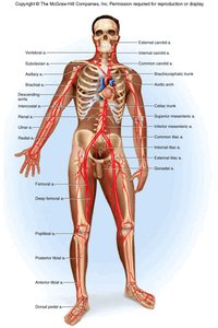
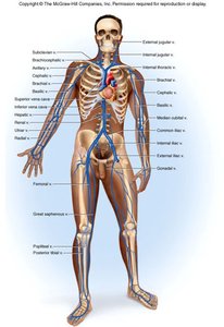
Coronary Circulation
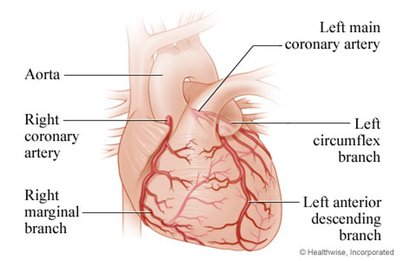
The heart has its own blood supply via the coronary arteries, which branch from the aorta and encircle the heart to provide oxygen and nutrients to cardiac muscle.
Left main coronary artery branches into the left anterior descending branch and left circumflex branch.
Right coronary artery gives rise to the right marginal branch.
Summary Table: Types of Blood Vessels and Their Functions
Vessel Type | Structure | Function | Example Location |
|---|---|---|---|
Artery | Thick, muscular, elastic | Carry blood away from heart | Aorta, femoral artery |
Arteriole | Small, muscular | Regulate blood flow to capillaries | Within organs |
Capillary | Single layer endothelium | Exchange of gases, nutrients, waste | All tissues |
Venule | Thin-walled | Collect blood from capillaries | Postcapillary beds |
Vein | Thin, less muscular, valves | Return blood to heart | Vena cava, jugular vein |
Additional info: Students should be able to identify the major arteries and veins, know which organs they supply or drain, and understand the functional significance of each vessel type.
