 Back
BackBlood Vessels: Structure, Function, and Clinical Correlations
Study Guide - Smart Notes
 Tailored notes based on your materials, expanded with key definitions, examples, and context.
Blood Vessels: Structure and Function
Overview of Blood Vessel Types
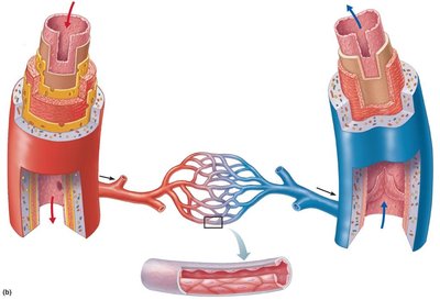
The human circulatory system is composed of three main types of blood vessels: arteries, veins, and capillaries. Each type has a unique structure and function, allowing for efficient transport of blood throughout the body.
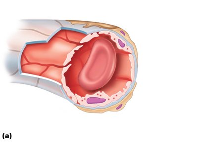
Arteries carry blood away from the heart under high pressure and have thick, muscular walls.
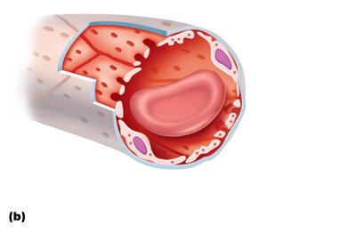
Veins return blood to the heart under lower pressure and often contain valves to prevent backflow.
Capillaries are microscopic vessels where exchange of gases, nutrients, and wastes occurs between blood and tissues.
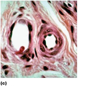
Histological Structure of Blood Vessels
Blood vessels are composed of three tunics (layers):
Tunica intima: The innermost layer, consisting of endothelium and a subendothelial layer.
Tunica media: The middle layer, primarily smooth muscle and elastic fibers, responsible for vasoconstriction and vasodilation.
Tunica externa (adventitia): The outermost layer, composed of collagen fibers that protect and anchor the vessel.
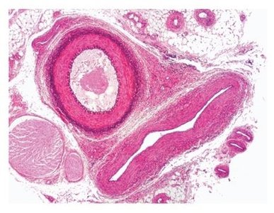
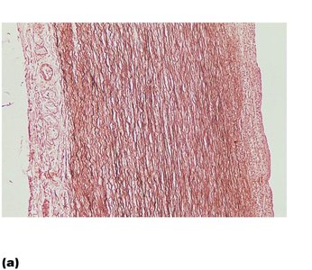
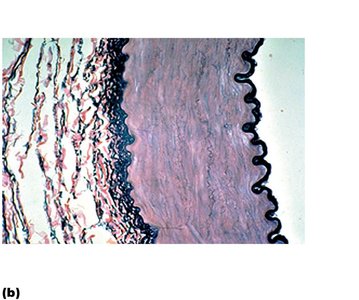
Comparison of Arterial Wall Structure
Arteries are classified based on the composition of their walls:
Elastic arteries (e.g., aorta): Large, thick-walled arteries with abundant elastin to withstand high pressure.
Muscular arteries: Distribute blood to specific organs; have a thick tunica media with more smooth muscle.
Arterioles: Smallest arteries, regulate blood flow into capillary beds via vasoconstriction and vasodilation.
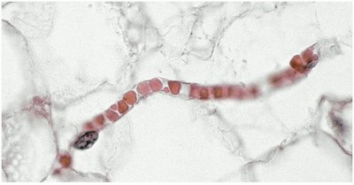
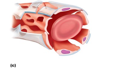
Capillaries: Structure and Types
General Structure and Function
Capillaries are the smallest blood vessels, consisting of a single layer of endothelial cells and a basement membrane. They facilitate the exchange of gases, nutrients, and waste products between blood and tissues.
Types of Capillaries
Continuous capillaries: Least permeable, most common; found in skin, muscle, and the brain. Characterized by tight junctions and intercellular clefts.
Fenestrated capillaries: Have pores (fenestrations) that increase permeability; found in kidneys, small intestine, and endocrine glands.
Sinusoid capillaries: Most permeable, with large gaps and an incomplete basement membrane; found in liver, bone marrow, and spleen.
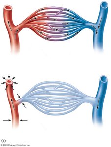
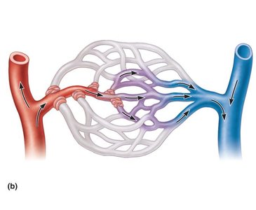
Capillary Beds and Microcirculation
Organization of Capillary Beds
Capillary beds are networks of capillaries supplied by arterioles and drained by venules. Blood flow through capillary beds is regulated by precapillary sphincters, which open or close to direct blood flow according to tissue needs.
When sphincters are open, blood flows through true capillaries for exchange.
When sphincters are closed, blood bypasses the capillary bed via a vascular shunt (metarteriole-thoroughfare channel).
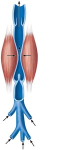
Venous System: Structure and Function
Veins and Venous Valves
Veins have thinner walls and larger lumens than arteries. They contain valves, especially in the limbs, to prevent backflow of blood. The movement of blood in veins is aided by skeletal muscle contraction and changes in thoracic pressure during breathing.
Venous valves: Prevent retrograde flow of blood.
Skeletal muscle pump: Muscle contractions compress veins, propelling blood toward the heart.
Major Circulatory Pathways
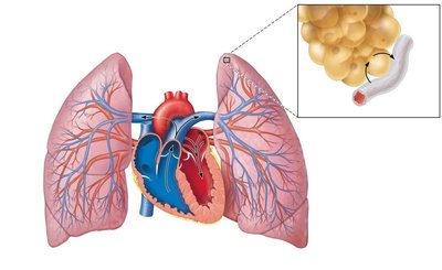
Pulmonary Circulation
Pulmonary circulation carries deoxygenated blood from the right ventricle to the lungs and returns oxygenated blood to the left atrium. Gas exchange occurs in the pulmonary capillaries surrounding alveoli.
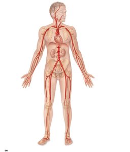
Systemic Circulation: Major Arteries and Veins
The systemic circulation delivers oxygenated blood from the left ventricle to all body tissues and returns deoxygenated blood to the right atrium. Major arteries and veins are named according to the regions and organs they supply or drain.
Major arteries: Aorta, carotid arteries, subclavian arteries, mesenteric arteries, iliac arteries, femoral arteries, etc.
Major veins: Superior and inferior vena cava, jugular veins, subclavian veins, hepatic veins, renal veins, iliac veins, femoral veins, etc.
Clinical Correlations and Scenarios
Deep Vein Thrombosis (DVT) Scenario
Case: A patient presents with unilateral swelling, pain, warmth, and redness in the calf after a long flight. These are classic signs of deep vein thrombosis (DVT), a condition where a blood clot forms in a deep vein, often in the legs. DVT is dangerous due to the risk of pulmonary embolism if the clot dislodges and travels to the lungs.
Key signs: Swelling, pain, warmth, redness, history of immobility.
Clinical importance: Prompt diagnosis and treatment are essential to prevent life-threatening complications.
Aortic Aneurysm Scenario
Case: An elderly male with a history of smoking and hypertension is found to have a dilation of the artery above the heart (likely the aorta). This is characteristic of an aortic aneurysm, a localized dilation of the aorta that can rupture if not managed.
Risk factors: Smoking, hypertension, age, male sex.
Diagnosis: Imaging reveals dilation; surgical intervention is considered if the risk of rupture outweighs surgical risks.
Repair approaches: Open surgical repair or endovascular stent grafting, depending on location and patient factors.
Summary Table: Comparison of Blood Vessel Types
Vessel Type | Wall Structure | Function | Key Features |
|---|---|---|---|
Artery | Thick tunica media, elastic fibers | Carry blood away from heart | High pressure, no valves |
Vein | Thin walls, large lumen | Return blood to heart | Low pressure, valves present |
Capillary | Single endothelial layer | Exchange of substances | Smallest vessels, permeable walls |
Key Terms and Concepts
Vasa vasorum: Small vessels that supply blood to the walls of large arteries and veins.
Lumen: The central blood-containing space of a vessel.
Precapillary sphincter: A ring of smooth muscle that regulates blood flow into capillaries.
Venous valve: A structure in veins that prevents backflow of blood.
Hepatic portal system: A specialized circulatory route that carries nutrient-rich blood from the digestive organs to the liver for processing.
Equations and Formulas
Blood Flow (F): Where is the pressure difference and is resistance.
Poiseuille's Law (for resistance): Where is blood viscosity, is vessel length, and is vessel radius.
Clinical Application and Exam Readiness
Understanding vessel structure is essential for interpreting clinical signs such as bruising, varicose veins, and vascular emergencies.
Knowledge of major arteries and veins is critical for procedures like IV insertion, blood draws, and interpreting imaging.
Clinical scenarios often test reasoning about blood flow, vessel pathology, and emergency management.
