 Back
BackBlood Vessels: Structure, Function, and Major Pathways
Study Guide - Smart Notes
 Tailored notes based on your materials, expanded with key definitions, examples, and context.
Blood Vessels: Structure, Function, and Major Pathways
Overview and Interesting Facts
Blood vessels are essential components of the circulatory system, responsible for transporting blood throughout the body. They deliver oxygen and nutrients to tissues and remove waste products, maintaining homeostasis and supporting tissue metabolism.
Total Length: The human body contains approximately 60,000 miles of blood vessels.
Capillaries: There are about 10 billion capillaries, facilitating exchange between blood and tissues.
Global Comparison: If laid end-to-end, blood vessels could wrap around the Earth's equator 2.5 times.
Function: Blood vessels deliver oxygen and nutrients to tissues and remove waste at a rate that matches tissue metabolism.

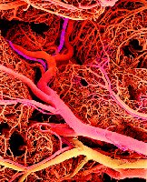
Anatomy of Blood Vessels
Blood vessels are classified based on their function and direction of blood flow:
Arteries: Carry blood away from the heart.
Veins: Carry blood back to the heart.
Capillaries: Connect the smallest arteries to the smallest veins, facilitating exchange.
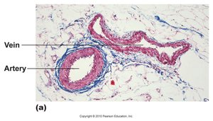
Structure of Blood Vessels
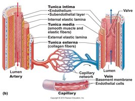
Blood vessels have three main layers, each with distinct functions:
Tunica intima: Innermost layer, composed of endothelium and subendothelial layer.
Tunica media: Middle layer, contains smooth muscle and elastic fibers, responsible for vasoconstriction and vasodilation.
Tunica externa: Outermost layer, composed of collagen fibers for structural support.
Valves: Present in veins to prevent backflow of blood.
Arteries
Arteries are resistance vessels, designed to withstand high blood pressure generated by ventricular contractions. They are muscular and retain a round shape when empty.
Types of Arteries:
Conducting (Elastic) Arteries: 2.5–1.0 cm diameter; e.g., aorta, pulmonary, common carotid. Expand and relax to maintain blood pressure.
Distributing (Muscular) Arteries: 1.0 cm–0.3 mm diameter; e.g., femoral, brachial, renal, splenic. Distribute blood to organs.
Resistance Arteries: 0.3 mm–10 μm diameter; arterioles. Control the amount of blood delivered to organs.
Arterial Sense Organs
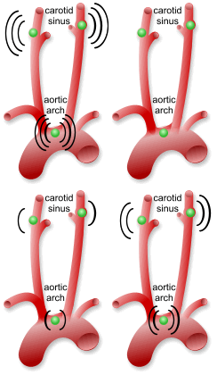
Arterial sense organs monitor blood pressure and chemical composition:
Baroreceptors: Located in the aortic arch and carotid arteries; detect changes in blood pressure and signal the heart to adjust heart rate.
Chemoreceptors: Sensitive to pH, CO2, and O2; located in the aortic arch and carotids; important for respiratory control.
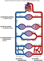
Portal system: Blood flows through two consecutive capillary networks before returning to the heart.
Arteriovenous shunt: Artery flows directly into a vein.
Venous anastomosis: Alternate drainage of organs; most common, blockage less serious.
Arterial anastomosis: Collateral circulation, e.g., coronary arteries.
Major Systemic Arteries
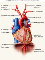
Systemic arteries supply oxygen and nutrients to all organs. The aorta is the largest artery, feeding oxygen-rich blood to all other systemic arteries.
Ascending aorta
Aortic arch
Thoracic (descending) aorta
Abdominal (descending) aorta
Left and right subclavian arteries
Left and right common carotid arteries
Brachiocephalic trunk
Aneurysm
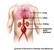
An aneurysm is a weak point in an artery or heart wall that can rupture. A dissecting aneurysm occurs when blood pools between vessel layers. Causes include congenital defects, trauma, atherosclerosis, and hypertension.
Coronary Arteries
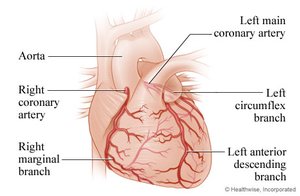
The coronary arteries branch from the ascending aorta and supply the heart muscle (myocardium).
Right Coronary Artery
Left Coronary Artery
Arteries of the Thorax
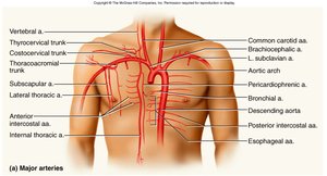
Thoracic arteries supply the viscera and body wall, including the common carotid arteries, brachiocephalic trunk, and vertebral artery.
Arteries of the Head and Neck
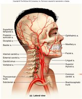
The common carotid artery branches into internal and external carotids. The external carotid supplies most external head structures, while the internal carotid supplies the brain and internal organs of the skull.
Arteries of the Upper Limb
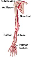
The subclavian artery passes between the clavicle and first rib, changing names as it moves through regions: subclavian → axillary → brachial → radial and ulnar. The brachial artery is used for blood pressure measurement, and the radial artery for pulse.
Major Branches of Abdominal Aorta
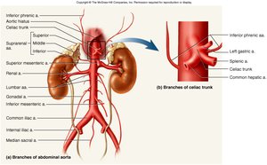
The abdominal aorta gives rise to several branches:
Renal artery
Common iliac artery
Internal iliac artery
Inferior and superior mesenteric arteries
Celiac trunk (first branch, with three subdivisions)
Gonadal artery
Arteries of the Lower Limb
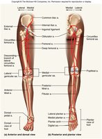
Branches to the lower limb arise from the external iliac branch of the common iliac artery, including the femoral, deep femoral, popliteal, posterior tibial, anterior tibial, and dorsal pedal arteries.
Major Systemic Veins
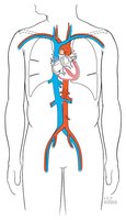
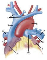
Veins return oxygen-poor blood to the heart. The superior and inferior vena cavae are the largest veins, delivering blood from the upper and lower body to the right atrium.
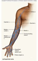
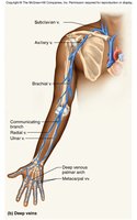
Brachiocephalic veins: Receive blood from the arm and head, delivering it to the superior vena cava.
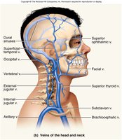
Veins of the head and neck: Internal jugular vein drains the brain; external jugular veins drain structures external to the skull; subclavian vein drains the upper limb.
Superficial and Deep Veins of Upper Limb
The cephalic vein drains the lateral side of the arm, the basilic vein drains the medial side, and they are connected by the median cubital vein. The subclavian vein lies deep to the clavicle, and the axillary vein runs through the armpit.
Inferior Vena Cava and Branches
The external and internal iliac veins join to become the common iliac vein. The internal iliac vein drains organs of the pelvic cavity.
Hepatic veins
Renal veins
Gonadal veins
Superficial and Deep Veins of Lower Limb
Major veins include the femoral vein, great saphenous vein, popliteal vein, posterior and anterior tibial veins, and external iliac vein.
Clinical Uses of Saphenous Vein
The great saphenous vein is often used in clinical procedures such as coronary artery bypass grafting due to its length and accessibility.
Type | Structure | Function | Location |
|---|---|---|---|
Artery | Thick, muscular, elastic | Carry blood away from heart | Systemic and pulmonary circuits |
Vein | Thin, less muscular, valves | Carry blood to heart | Systemic and pulmonary circuits |
Capillary | Single layer endothelium | Exchange of gases, nutrients, waste | Between arteries and veins |
Additional info: The notes have been expanded to include definitions, examples, and clinical context for each vessel type and major pathway, as well as a comparative table for clarity.
